Fig. 2.

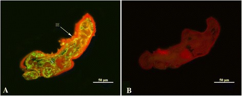
Immunolocalization of cofilin in sections of Sarcoptes scabiei. Panel a: staining with anti-cofilin as primary antibody; Panel b: control (no primary antibody). Annotation: S, splanchnic area; IE, epidermal integument

Immunolocalization of cofilin in sections of Sarcoptes scabiei. Panel a: staining with anti-cofilin as primary antibody; Panel b: control (no primary antibody). Annotation: S, splanchnic area; IE, epidermal integument