Fig. 6.

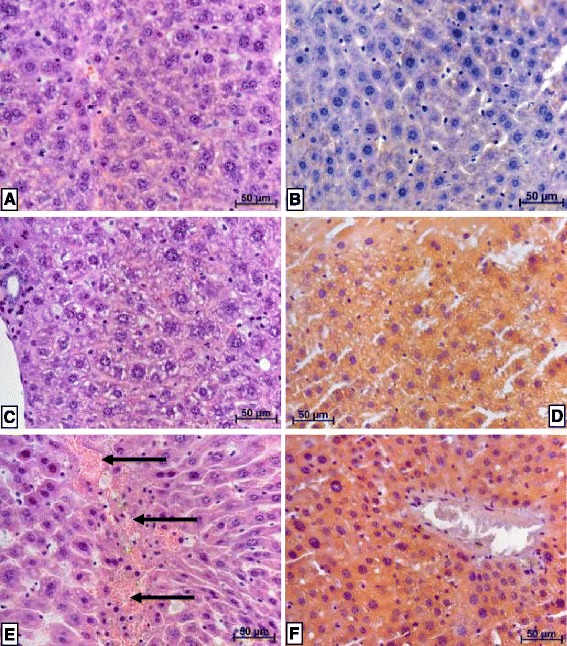
Lipid staining in liver of mice after 1 month of P-407 administration. H&E = hematoxylin and eosin staining, while S&H = Sudan III and hematoxylin staining. Magnification is represented by linear distance according to the scale on each histology slide (a) through (f). a Liver of control mice, H&E. b Liver of control mice, S&H. c Liver of mice 24 h after discontinuation of P-407 administration for 1 month. Mild vacuolar dystrophy of hepatocytes, together with necrosis of some hepatocytes, H&E. d Liver of mice 24 h after discontinuation of P-407 administration for 1 month. All hepatocytes are filled with small and medium-sized lipid droplets, thus displaying significant lipid dystrophy, S&H. e Liver of mice at 4 days after discontinuation of P-407 administration for 1 month. Hepatocytes are significantly enlarged and homogenously stained. There is diffuse leukocyte infiltration visible. The portion of hepatocytes demonstrating necrosis appear to have incorporated erythrocytes (arrows), H&E. f Liver of mice 4 days after discontinuation of P-407 administration for 1 month. All hepatocytes are filled with small lipid droplets, thus displaying significant lipid dystrophy, S&H
