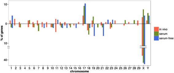
Fig. 3.

Chromosome distribution of the differentially expressed genes between male and female embryos. The bars represent the percentage of genes differentially expressed (FDR corrected p-value <0.05 and │FC│ ≥2) up- regulated (above) and down- regulated (below) in males vs. females belonging to each chromosome in each of the three conditions studied
