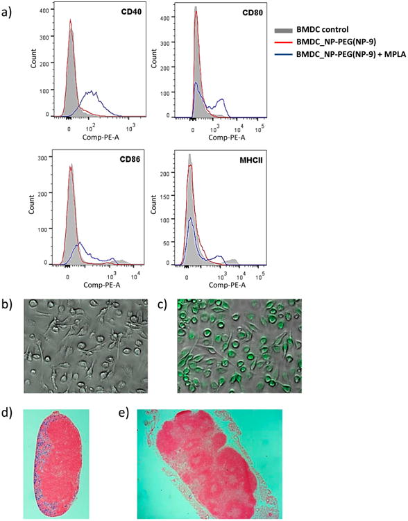
Figure 3.

(a) Flow cytometry results showing the expression of cellular markers of the activation state of BMDCs (CD40, CD80, CD86, and MHCII) after incubation with NP-PEG (NP-9) (red line) or NP-PEG (NP-9) + MPLA (blue line). Confocal images of BMDCs incubated with (b) PBS and (c) NP-9 (FITC) + MPLA. Histology of sections of an (d) axillary (local) lymph node and (e) inguinal (distant) lymph node stained by Prussian blue.
