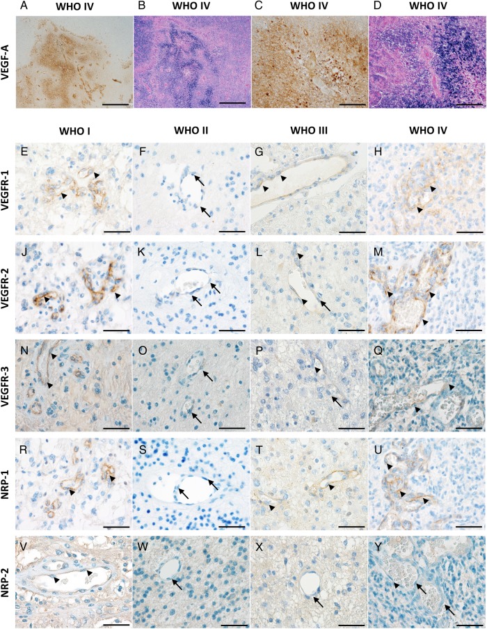
Fig. 1.
VEGF-A, VEGFR-1, -2, -3 and NRP-1 and -2 expression in human astrocytomas by in-situ hybridization ISH) and immunohistochemistry (IHC). VEGF-A: (A) IHC-staining of a glioblastoma with high VEGF-A levels on tumor cells and tumor vessels. (B) Corresponding ISH of the same area on a serial section showing similar mRNA signals. (C and D) Higher magnification of corresponding areas of the same tumor. Scale bars (A) and (B) = 1000 µm, (C) and (D) = 200 µm. VEGFR-1: (E) Pilocytic WHO grade I astrocytoma showing moderate-to-strong expression of VEGFR-1 on endothelial cells (arrowheads). (F) WHO grade II astrocytoma exhibiting VEGFR-1 negative vessels (arrows). (G) WHO grade III astrocytoma with weak endothelial staining for VEGFR-1 (arrowheads). (H) Representative vital tumor center of a glioblastoma with VEGFR-1 positive endothelial cells (arrow-heads). VEGFR-2: (J) Pilocytic astrocytoma WHO grade I showing strong expression of VEGFR-2 on endothelial cells (arrowheads). (K) WHO grade II astrocytoma with VEGFR-2 negative vessels (arrows). (L) WHO grade III astrocytoma exhibiting weak endothelial staining for VEGFR-2 (arrowheads) and negative endothelial cells (arrow). (M) Representative vital tumor center of a glioblastoma with VEGFR-2 positive endothelial cells (arrowheads). VEGFR-3: (N) WHO grade I pilocytic astrocytoma showing moderate-to-strong VEGFR-3 expression on endothelial cells (arrowheads). (O) WHO grade II astrocytoma with VEGFR-3 negative vessels (arrows). (P) WHO grade III astrocytoma with weak endothelial staining for VEGFR-3 (arrowheads) and negative endothelial cells (arrow). (Q) Representative vital tumor center of a glioblastoma with VEGFR-3 moderately positive vessels (arrowheads). NRP-1: (R) WHO grade I pilocytic astrocytoma showing strong NRP-1 expression on tumor vessels (arrowheads). (S) WHO grade II astrocytoma with NRP-1 negative vessels (arrows). (T) WHO grade III astrocytomas with weak endothelial staining for NRP-1 (arrowheads). (U) Representative vital tumor center of a glioblastoma with high NRP-1 levels on tumor vessels (arrowheads). NRP-2: (V) WHO grade I pilocytic astrocytoma showing low NRP-2 expression on blood vessels (arrowheads). (W) WHO grade II and (X) WHO grade III astrocytomas with NRP-2 negative vessels (arrows). (Y) Vital tumor center of a glioblastoma with low vascular NRP-2 levels on some of the endothelial cells (arrowhead) while others remain negative (arrows). Scale bars = 50 µm.

