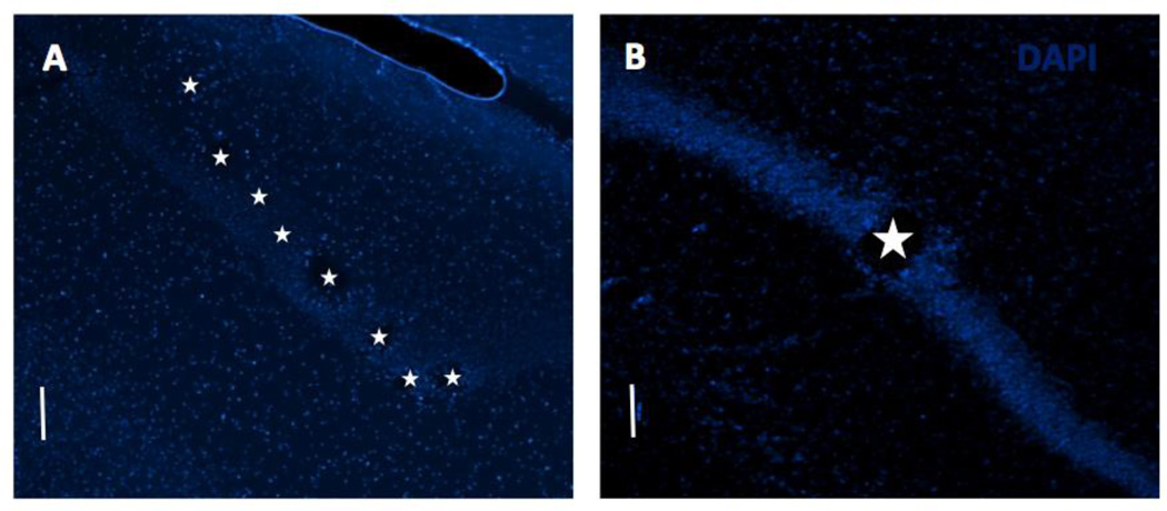
Figure 1.
DAPI stained horizontal sections through dorsal hippocampus to confirm implantation site. Tissue damage due to electrode implantation can be seen along the pyramidal cell layer in the (A) MEA implanted and (B) macroelectrode implanted cases. Stars mark the electrode locations. Scale bar: 100 µm.

