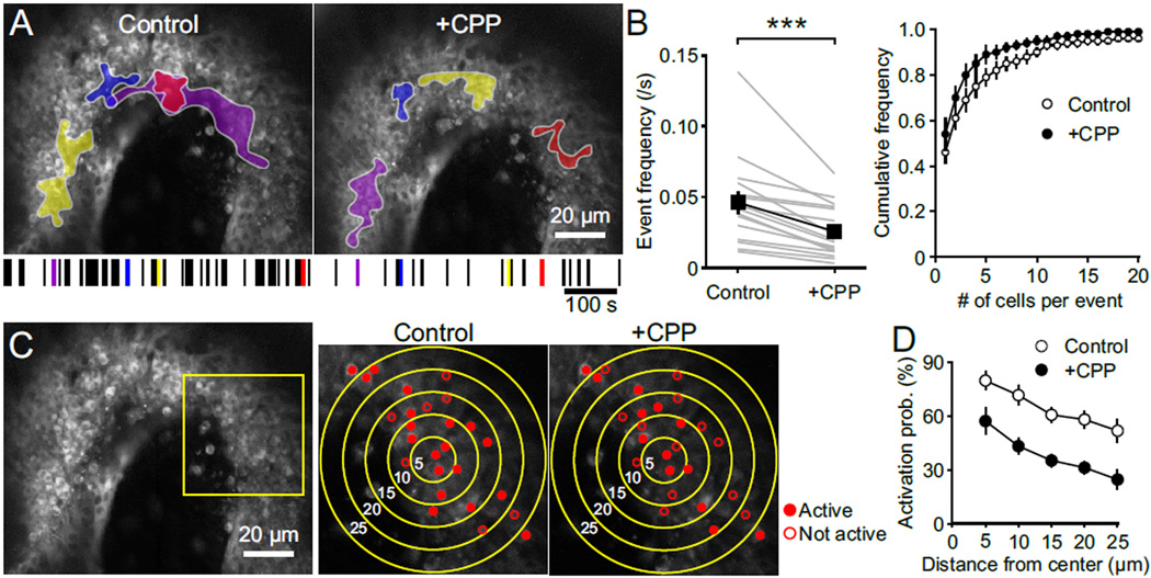
Figure 7. NMDAR Activation Promotes Coincidental Activation of Neighboring Spiral Ganglion Neurons.
(A) Maximum intensity projections of fluorescence changes in the spiral ganglion recorded for 300 s in control and + CPP overlaid with maximum activated area of four spontaneous events for both conditions. A raster plot of the timing of spontaneous events in the spiral ganglion is shown at the bottom, with the four examples shown above highlighted by their corresponding colors.
(B) Left: Plot of the correlated spontaneous event frequency in control and + CPP. n = 16 cochleae; paired-sample t test; ***, p < 0.001. Data show average values from each cochlea (grey) and mean ± SEM for all cochleae (black). Right: Cumulative frequency distribution of the number of activated SGNs per spontaneous event in control (open) and + CPP (filled) conditions. n = 16 cochleae; two-sample Kolmogorov–Smirnov test; p = 0.03. Data show mean ± SEM for all cochleae.
(C) Maximum intensity projection image of the spinal ganglion (left) showing active (filled) and not active (open) SGNs during single spontaneous events in control and + CPP. Higher magnification images at right were taken from area outlined by yellow square in left panel. Concentric circles indicate 5, 10, 15, 20 and 25 µm away from the location of the first responding SGN.
(D) Plot of activation probability of SGNs versus the distance from location of the first responding SGN in control (open) and + CPP (filled). n = 7 cochleae; two-way repeated measures ANOVA followed by Bonferroni test; p < 0.001. Data show mean ± SEM for all cochleae.
See also Movie S3.

