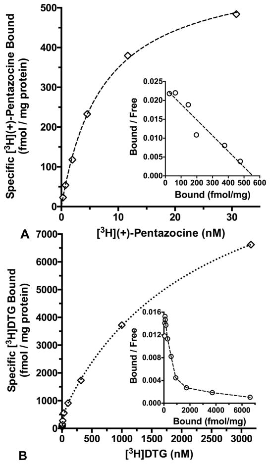
Fig. 7.
Panel A: Saturation binding of [3H](+)-pentazocine to CD-1® mouse brain membranes at 37 ºC with a 180 min incubation and haloperidol (1.0 μM) to define non-specific binding. Panel B: Saturation isotherm for [3H]DTG binding to CD-1® mouse brain membranes at 25 ºC with a 60 min incubation, 500 nM (+)-pentazocine to mask σ1 binding, and haloperidol (10.0 μM) to define non-specific binding. Open diamonds show specific binding, while open circles depict the inset Rosenthal plots. Data are representative experiments performed in duplicate, and replicated 4 – 6 times.

