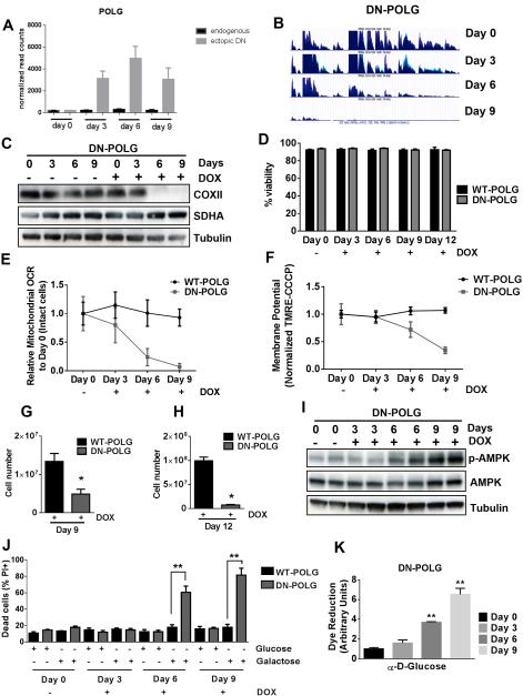
Figure 1. Inducible expression of DN-POLG in HEK293 cells diminishes mitochondrial respiration, mitochondrial membrane potential, and cell proliferation.
(A) Expression of the ectopic DN-POLG increases in cells treated with doxycycline (10 ng/ml) for 3, 6 and 9 days. Mean ± SEM (n=3).
(B) RNA-seq analysis of mitochondrial transcripts in three independent samples of DN-POLG cells untreated or treated with doxycycline (10 ng/ml) for 3, 6 and 9 days.
(C) Representative western blots of the expression of COXII (mtDNA encoded) and SDHA (nuclear encoded) in DN-POLG cells untreated or treated with doxycycline (10 ng/ml) for 3, 6 and 9 days from 3 independent experiments.
(D) WT and DN-POLG cells were left untreated or treated with doxycycline (10 ng/ml) for 3, 6, 9 for 12 days and viability was measured. Mean ± SEM (n=4).
(E-F) Relative mitochondrial oxygen consumption rate (OCR) (E) and mitochondrial membrane potential (F) of intact WT and DN-POLG cells untreated or treated with doxycycline (10 ng/ml) for 3, 6 and 9 days. Mean ± SEM (n=4).
(G-H) WT and DN-POLG cells were treated with doxycycline (10 ng/ml) and cell number was assessed at days 9 (G) and 12 (H). Mean ± SEM (n=3).
(I) Western blot analysis of p-AMPK, AMPK and Tubulin proteins in DN-POLG cells untreated or treated with doxycycline (10 ng/ml) for 3, 6 and 9 days.
(J) WT and DN-POLG untreated or treated with doxycycline (10 ng/ml) for 3, 6 and 9 days were grown in media containing 10 mM glucose or 10 mM galactose for 48h and assessed for cell death. Mean ± SEM (n=3).
(K) Glucose oxidation by DN-POLG cells untreated or treated with doxycycline (10 ng/ml) for 3, 6 and 9 days. Mean ± SEM (n=3).
* indicates significance p < 0.05. ** indicates significance p <0.01 throughout figure 1. See also Figure S1.

