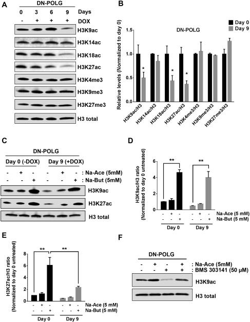
Figure 2. Inducible expression of DN-POLG in HEK293 cells diminishes specific histone H3 acetylation marks.
(A-B) Western blot analysis and quantification of histone 3 acetylation and methylation marks in DN-POLG cells untreated or treated with doxycycline (10 ng/ml) for 3, 6 and 9 days. Mean ± SEM (n=3).
(C) Acetylation state of H3K9 and H3K27 after treating DN-POLG cells at days 0 and 8 of doxycycline (10 ng/ml) treatment with 5 mM sodium acetate (Na-Ace) or 5 mM sodium butyrate (Na-But) for 24h.
(D-E) Quantification of the western blots assessing the acetylation of H3K9 (D) and H2K27
(E) of DN-POLG cells at days 0 and 8 of doxycycline (10 ng/ml) treatment in the presence or absence of 5 mM sodium acetate or sodium butyrate for 24h. Mean ± SEM (n=3).
(F) Acetylation state of H3K9 after treating DN-POLG cells with 5 mM sodium acetate (Na-Ace), 50 μM BMS 303141 (ACL inhibitor) and the combination of both treatments for 48h.
* indicates significance p < 0.05. ** indicates significance p <0.01 throughout Figure 2. See also Figure S2.

