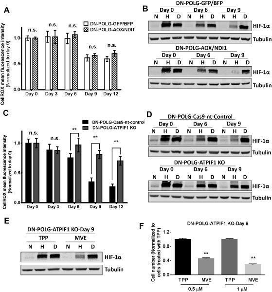
Figure 7. Mitochondrial membrane potential dependent ROS is essential for hypoxic stabilization of HIF-1α protein and cell proliferation.
(A) ROS levels were measured in DN-POLG-GFP/BFP and DN-POLG-AOX/NDI1 cells untreated or treated with doxycycline (10 ng/ml) for 3, 6, 9 and 12 days. Mean ± SEM (n=3).
(B) Representative western blot from 3 independent experiments of DN-POLG-GFP/BFP and DN-POLG-AOX/NDI1 cells untreated or treated with doxycycline (10 ng/ml) for 6 and 9 days and placed in normoxia (21% O2), hypoxia (1.5% O2) or treated with 1 mM DMOG (21% O2) for 4 hours. Hypoxic induction of HIF-1α was analyzed by western blot.
(C) ROS levels were measured in DN-POLG-Cas9-nt-control and DN-POLG-ATPIF1 KO cells untreated or treated with doxycycline (10 ng/ml) for 3, 6, 9 and 12 days. Mean ± SEM (n=3).
(D) Representative western blot from 3 independent experiments of DN-POLG-Cas9-nt-control and DN-POLG-ATPIF1 KO cells untreated or treated with doxycycline (10 ng/ml) for 6 and 9 days and placed in normoxia (21% O2), hypoxia (1.5% O2) or treated with 1 mM DMOG (21% O2) for 4 hours. Hypoxic stabilization of HIF-1α was analyzed by western blot.
(E) Representative western blot from 3 independent experiments of DN-POLG-ATPIF1 KO cells treated with doxycycline (10 ng/ml) for 9 days pretreated with TPP or MVE for 2 hours and subsequently placed in normoxia (21% O2), hypoxia (1.5% O2) or treated with 1 mM DMOG (21% O2) for 4 hours. Hypoxic stabilization of HIF-1α was analyzed by western blot.
(F) DN-POLG-ATPIF1 KO cells treated with doxycycline (10 ng/ml) for 9 days were exposed to 0.5 μM or 1 μM TPP or MVE for the last 3 days of doxycycline treatment and cell number was assessed. Mean ± SEM (n=3).
** Indicates significance p <0.01 throughout figure 7. See also Figure S7.

