
Figure 3. Classical alpha RGCs and their “mini” counterparts.
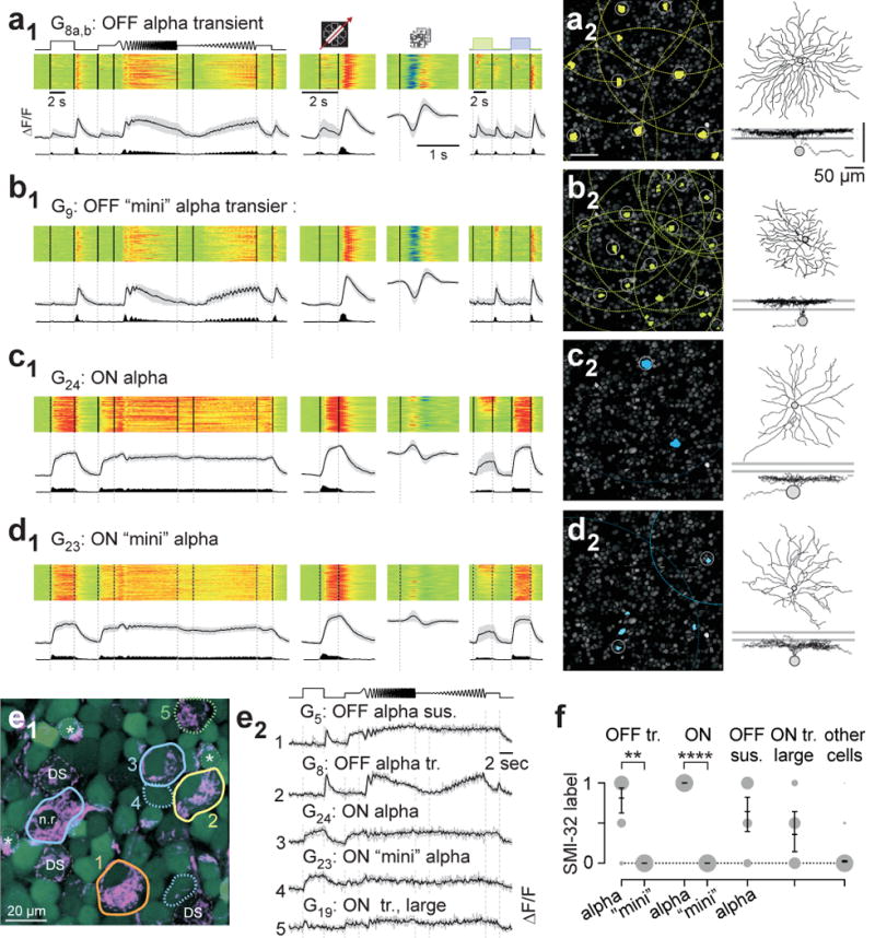
a, functional “fingerprint” of classical transient OFF alpha cells (G8a,b). Light-evoked Ca2+ responses of n=80 cells (a1): heat-maps (top) of individual responses, response averages and firing rates estimated from Ca2+ signals below (cf. Extended Data Fig. E1). a2, left, G8a,b somata (yellow) and receptive fields (RFs, dotted) indicated in example experiment (cf. Fig. 1a2). Grey circles mark cells with RFs above quality criterion (Methods). Sample morphology of a G8a,b cell filled after electrical single-cell recording (a2, right). For details, see SI Data 28. b1,2, G9 RGCs (n=68), dubbed transient OFF mini alpha because of their similarity in light response to G8a,b RGCs (cf. a1,2). c1,2, G24 RGCs (n=44), identified as classical ON alpha. d1,2, G23 RGCs (n=113), dubbed ON mini alpha (cf. c1,2). Mini alphas have smaller RF diameters than classical alphas (median in μm, 95% confidence interval): G9,280 (270–293) versus G8, 306 (294–315) with p=0.01232), and G23, 236 (218–256) versus G24, 319 (290–352) with p=0.00026 (rank-sum test). e, overlay of OGB-1-stained cells (green) and SMI-32 (magenta). SMI-32-positive RGCs include classical alphas (e1, solid contours; n.r. indicates a non-responsive cell), one large-soma non-alpha cell (green, dotted contour) as well as weakly-labeled ON-OFF DS cells (dashed contours) and starburst-ACs (asterisks). Mini alpha cells (blue, dotted contours) are SMI-32-negative. Chirp-evoked Ca2+ responses (e2) for 5 cells in (e1). f, SMI-32 statistics (OFF tr.: alpha, n=16, mini, n=3; ON: alpha, n=6, mini, n=15; OFF sus. alpha, n=14; ON tr. large, n=7; other, n=957; means with 95% confidence intervals, **, p≤0.01; ****, p≤0.0001, logistic regression).