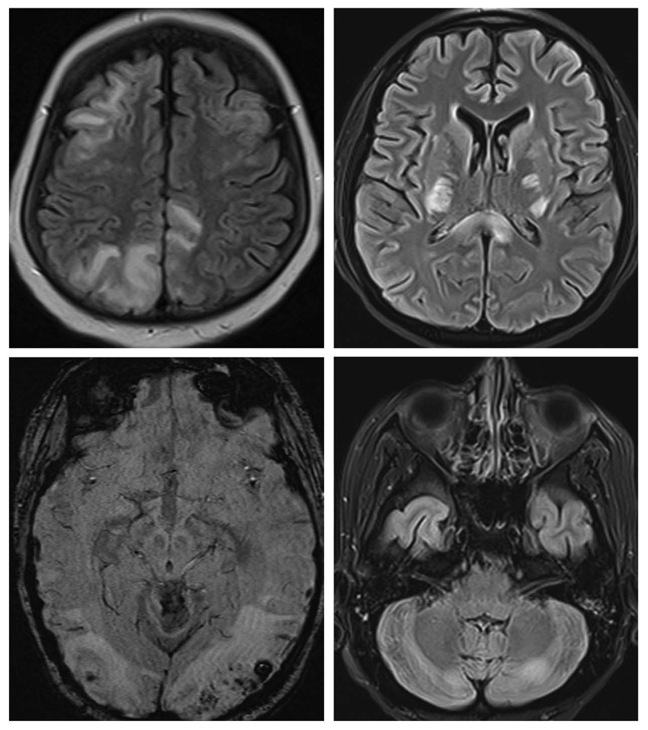
Figure 1.
FLAIR images showing edema involving white matter and cortex in occipital, parietal and frontal lobes in top left; central variant of PRES with edema in internal capsule, corpus callosum and basal ganglia in top right; and cerebellar grey and white matter in bottom right. Susceptibility weight image showing multiple micro hemorrhages in bilateral occipital area in the bottom left image.

