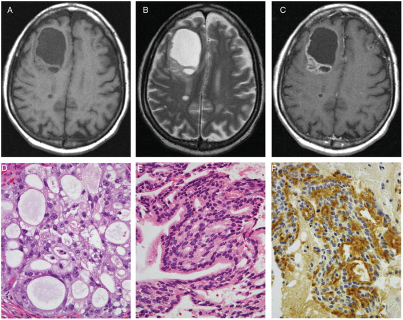
Fig 4.

The precontrast T1-weighted (A), T2-weighted (B), and postcontrast T1-weighted images (C) demonstrate a large predominantly cystic/necrotic right frontal mass with a heterogeneously enhancing solid component. Two smaller ring-enhancing lesions are present in the posterior right frontal lobe and superficial left frontal lobe. The dominant mass has been resected and exhibits confirmation from pathology findings as originating in prostatic adenocarcinoma. Hematoxylin and eosin stained sections of both the primary (D) and metastatic (E) tumor exhibit similar cribriform architecture. Immunostaining for prostate specific antigen (PSA) is positive in the metastasis (F).
