
Figure 1.
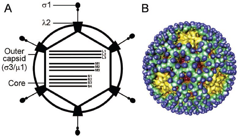
The reovirus virion. A) Schematic of a reovirus virion. Reovirus virions are composed of two concentric protein shells, outer capsid and core. The core contains the viral genome, which consists of 10 segments of double-stranded RNA. B) Cryo-EM image reconstruction of a reovirus virion at 23 Å resolution. Note the finger-like projections of σ3 (blue) that sit atop a layer of μ1 (green). The λ2 protein (yellow) forms a pentamer at each of the virion fivefold symmetry axes. Figure modified from: Nason E et al, J Virol 75:6625-6634; ©2001 with permission from the American Society for Microbiology.74