
Figure 4.
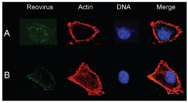
β1 integrin enhances reovirus entry into cells. (A) GD25β1A (β1 +/+) and (B) GD25 (β1 -/-) cells were chilled, adsorbed with T1L virions, and incubated at 4°C for 1 h. Nonadherent virus was removed, warm medium was added, and cells were incubated at 37°C for 30 min. Cells were fixed, stained for reovirus (green), actin (red), and DNA (blue), and imaged using confocal immunofluorescence microscopy. Representative digital fluorescence images of the same field are shown in each row. Figure modified from: Maginnis MS et al, J Virol 80:2760-2770; ©2006 with permission from the american Society for Microbiology.40