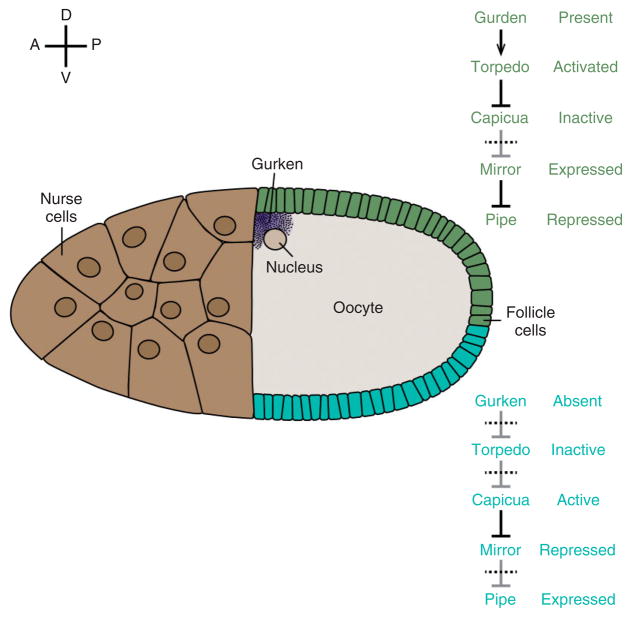
FIGURE 4.
Model for the ventrally restricted expression of pipe in the follicle cell layer. Schematic drawing of a stage 10 oocyte. pipe is expressed in the ventral region (blue) but repressed in the dorsal epithelium (green). Relevant effector molecules and their activation states in ventral and dorsal follicle cells are indicated at right.

