
FIGURE 5.
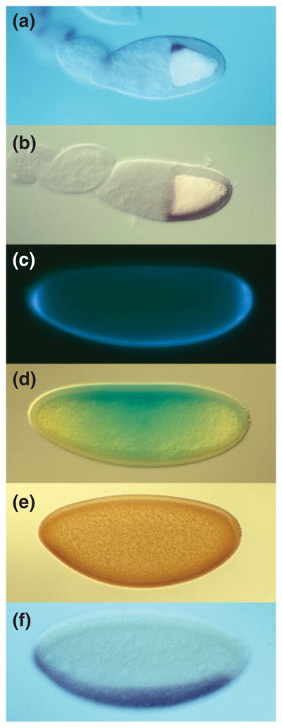
Sequential asymmetry along the DV axis of polarity from the oocyte to zygotic gene expression in the embryo. In all panels, anterior is to the left and ventral is down. (a, b) Stage 10 oocyte showing the dorsal anterior localization of gurken mRNA (a) and ventrally restricted expression of pipe mRNA (b). (c–e) Syncytial blastoderm embryos showing ventral localization of GD-GFP after injection into the pervitelline space (c), ventral-to-dorsal gradient of Cactus-LacZ degradation visualized by X-gal staining (d), and ventral-to-dorsal gradient of Dorsal nuclear localization visualized with anti-Dorsal antibody (e). (f) Cellular blastoderm embryo showing the expression domain of the mRNA encoding the Dorsal target gene twist in the presumptive mesoderm.