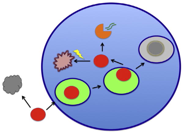
Figure 3. Infectivity decay in suspension and abortive events during attachment and after entry.
While virions are diffusing at random, they may lose their infectivity and become inert particles (gray) because of the exponential decay of their components; for enveloped viruses the decay of the envelope glycoproteins may often determine the infectivity half-life. The virion (red) is depicted as getting endocytosed (the endosomal lumen is bright green) and as it later either fuses with or penetrates from the endosome. In case of excessive delay before cytoplasmic entry, the endosome continues to the lysosomal compartment and the virion gets degraded (gray). In case of successful entry of the capsid into the cytoplasm, a productive uncoating (orange) may lead to genome (green curved double lines) release for transcription or, in competition with this, degradation of the whole core by the proteasome (yellow flash).

