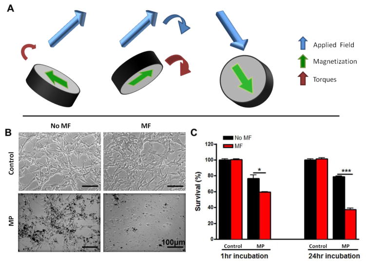
Figure 1. In vitro cell destruction using MPs under a rotating magnetic field (MF).
(A) Schematic of one of our MPs under a rotating MF. (B) Optical images of U87 glioma cells with (MF) and without (no MF) MF treatment (1 Tesla at 20 Hz for 30 min). Cells were treated with either growth media (control) or MPs at 50 particles per cell for 24 hours. Scale: 100 μm. (C) Quantification of the U87 cells viability after incubation with the MPs for 1 hour and 24 hours or not, and after MF treatment or not. Data are presented as Mean±SE. * p<0.05, *** p<0.001 (Student’s t test).

