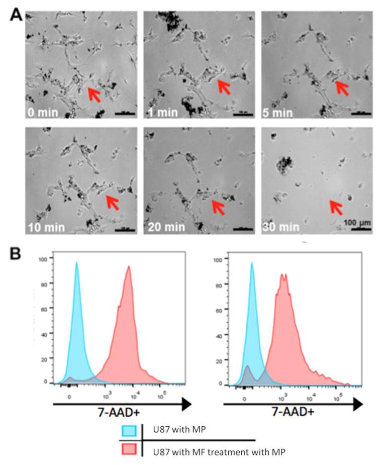
Figure 3. MPs compromise the membrane integrity of cancer cells under magnetic field.
(A) Optical images of nanomagnet-loaded U87 cells after MF treatment. Cells were incubated with MPs at 50 particles per cell for 24 hours. Images were taken from 0 min to 30 min post magnetic field treatment (1Tesla, 20Hz). The same area was monitored as indicated as the red arrow. (B) Flow cytometry analysis of cells (10000 events) 5 min and 30 min after MF treatment. The cells compared with the controls that were incubated with particles show 89.3% and 87% 7-AAD staining respectively.

