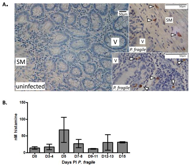
Figure 1. Plasmodium fragile-infected Rhesus macaques exhibit increased ileal mastocytosis and plasma histamine levels.
(A) Representative ileum sections from macaques infected with Plasmodium fragile immunostained with anti-tryptase and counter-stained with hematoxylin. Mast cells were detected in P. fragile-infected animals (arrowheads, n=4) but not in uninfected controls (n=4). SM denotes submucosa, V denotes villi. Bars equal 50 μm. (B) Mean histamine levels (+/− SEM) in plasma of P. fragile-infected macaques from panel A.

