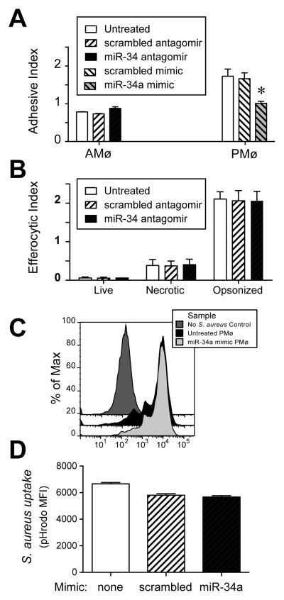
Figure 3. miR-34a negatively regulates binding to AC but not uptake of other targets.
A–D. Using RNAiMAX Lipofectamine and chamber slides, murine AMø were transfected with control or miR-34a-specific antagomirs (24 h incubation) and murine PMø were transfected with control or miR-34a-specific mimics (48 h incubation). A. AC adhesion by transfected AMø and PMø was assessed by histology after 15 min. Data are mean ± SEM of three independent experiments. B. Phagocytosis by transfected AMø was assessed by microscopy after 1.5 h exposure of AMø to live thymocytes, necrotic thymocytes, or opsonized SRBC. Data are mean ± SEM of at least three independent experiments per target cell type. C, D. Phagocytosis by transfected PMø of pHrodo-labeled, Ig-opsonized-S. aureus following 1 h exposure, then release and analysis by flow cytometry. C. Representative histogram showing pHrodo staining. D. Uptake as measured by pHrodo MFI; data are mean ± SEM from n=3 mice assayed individually in each of two independent experiments, p<0.05 by one-way ANOVA with Bonferroni post-hoc testing.

