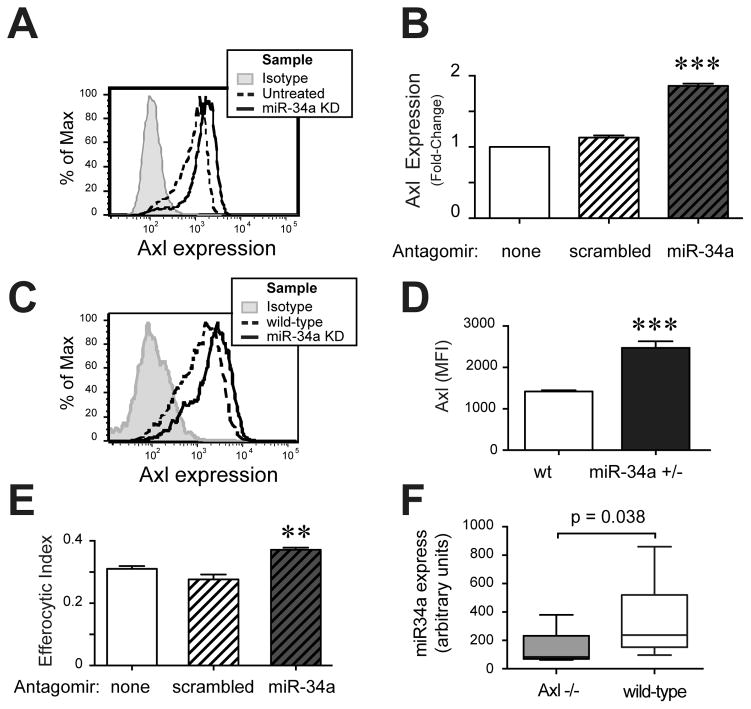
Figure 4. miR-34a regulates Mø expression of Axl, but altered Axl expression is not required for regulation of efferocytosis.
A, B. AMø from wt mice were transfected with control scrambled antagomirs (scrambled) or miR-34a-specific antagomirs (miR-34a KD) using RNAiMAX lipofectamine. Following knockdown, AMø were stained for surface expression of Axl and assayed by flow cytometry, gating AMø as CD45+ CD11c+ cells. A. Representative histograms; isotype, grey; untreated, dashed line; miR-34a+/− KD, solid line; scrambled antagomir-treated omitted for clarity. B. Data are expressed as fold change in MFI, relative to untreated AMø and are presented as mean ± SEM of as least four replicates in each of three independent experiments; ***, p<0.001 significantly different from both other groups, one-way ANOVA with Tukey post-hoc testing. C, D. BMDMø harvested from wt mice and miR-34a+/− mice were surface stained for Axl and analyzed by flow cytometry, gating on CD45+ cells. C. Representative histogram of Axl staining (isotype, grey; wt BMDMø, dashed line; miR-34a+/− BMDMø, solid line). D. Axl MFI; data are mean ± SEM of eight mice of each genotype in three independent experiments; ***, p<0.001, Mann-Whitney U test. E. BMDMø from Axl−/− mice were transfected with scrambled or miR-34a-specific antagomirs using RNAiMAX Lipofectamine, then exposed to AC for 90 min and assayed for efferocytosis by microscopy; data are mean ± SEM of duplicate or triplicate samples from three separate BMDMø cultures; **, p<0.01 by one-way ANOVA with Tukey post-hoc testing. F. miR-34a mRNA expression of BMDMø from Axl −/− mice (grary) or wild-type mice (white) was assayed by quantitative real-time PCR, relative to RNU6B; data are median, IQR and 95, 5% CI of eight independent bone marrow cultures assayed in four independent expreiments; Mann-Whitney U test.

