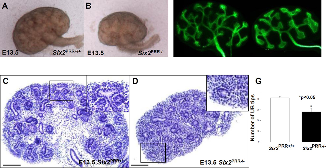
Figure 2. Nephron progenitor PRR is required for normal kidney development.
(AD) On E13.5, Six2PRR−/− kidneys are reduced in size (A, B) and have less developed epithelial structures on hematoxylin and eosin-stained sections cut in the longitudinal midplane (C, D). Insets in the upper right-hand corners of C and D show higher magnification photos of the respective boxed region. In control kidney (C), nascent vesicle and comma-shaped nephron are observed on ventral surface of branching UB. Mutant kidney is smaller and has no apparent developing nephron structures adjacent to the UB (D, inset). (E–G) The number of UB tips (green staining, anti-pancytokeratin antibody) is reduced in E13.5 mutant compared to control kidneys. (C,D) Scale bars= 100 µm.

