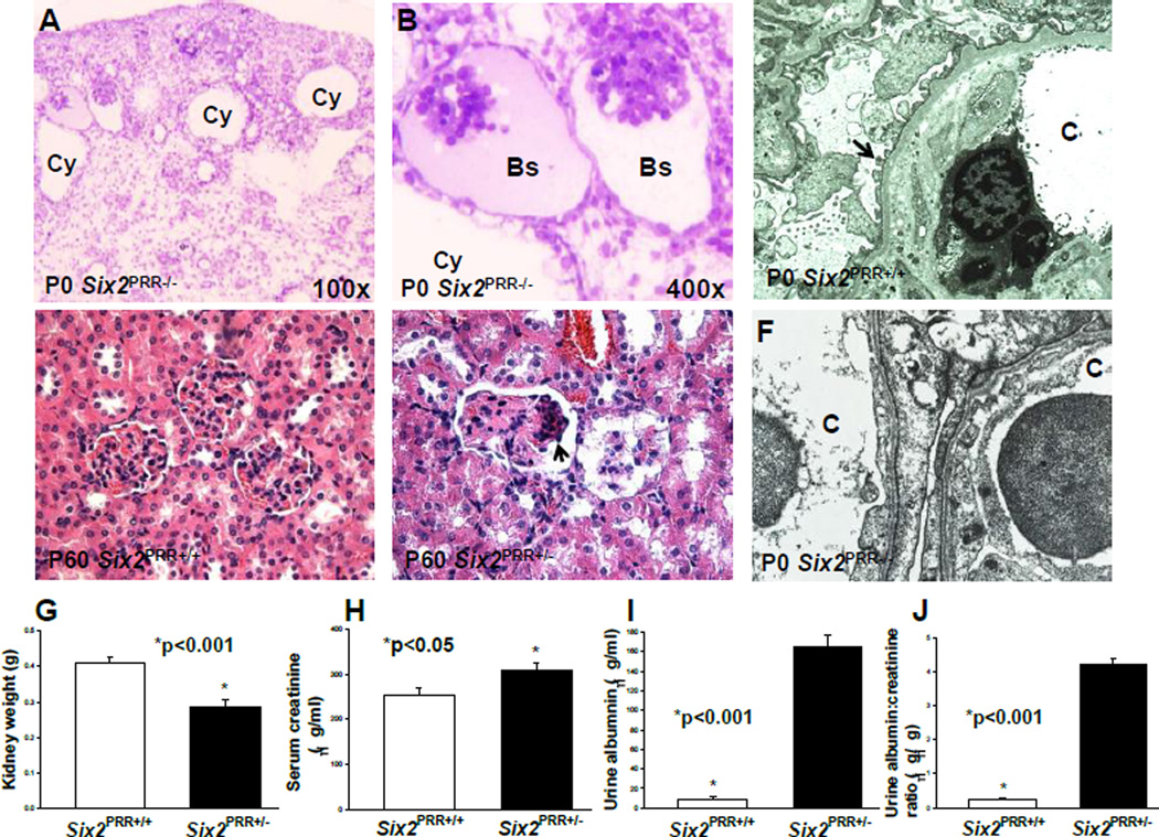
Figure 7. Nephron progenitorPRRis required for normal podocyte ultrastructure and kidney function.
(A,B) Toluidine blue-stained P0 Six2PRR−/− kidney sections show cystically dilated tubules (Cy) and collapsed glomeruli with an enlarged Bowman’s space (Bs). (C) Electron micrograph of P0 Six2PRR+/+ kidney section shows normally appearing podocyte foot processes (arrow). (F) P0 Six2PRR−/− kidney section shows fusion and effacement of podocyte foot processes in mutants (arrows); C- capillary lumen (original magnification x20,000). (D–E) Hematoxylin and eosin staining of 2 months-old kidney tissues shows normally appearing glomeruli in Six2PRR+/+ mice (D) and focal glomerulosclerosis (arrows) in Six2PRR+/− mice (E). (G) Kidney weight is reduced in Six2PRR+/− compared with Six2PRR+/+ mice at 2 months of age. (H–J) Six2PRR+/− mice have significantly increased levels of serum creatinine, urinary albumin and urinary albumin-to-creatinine ratios at 2 months of age.

