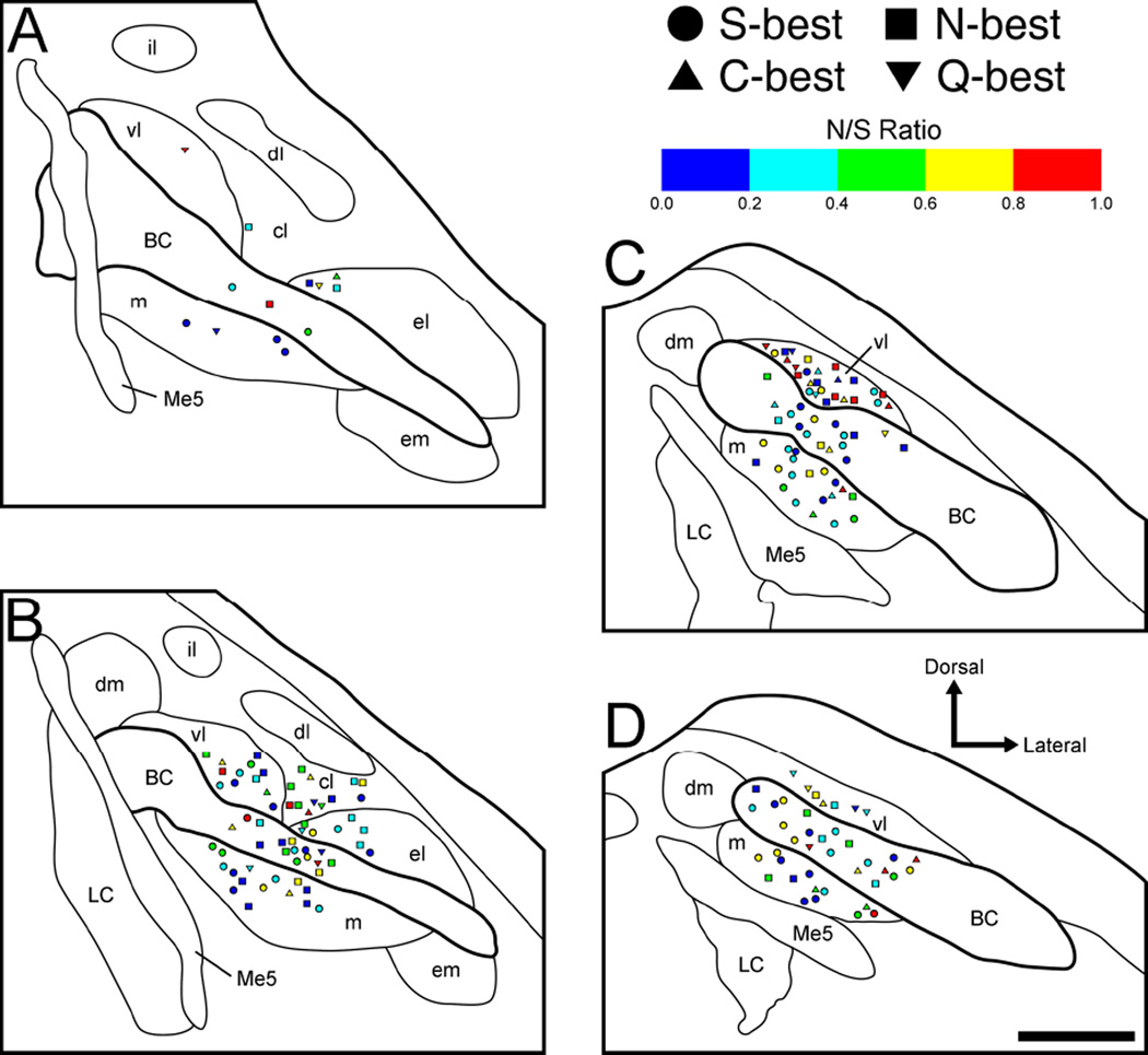
Figure 9.
Anatomical reconstruction of 178 recording sites in the right PbN in terms of noise-to-signal (N/S) ratio and best stimulus category. A–D: coronal sections are arranged rostral to caudal and +50, −100, −250, and −400 µm separated from the caudal end of the cuneiform nucleus, respectively. S-best units; squares, N-best units; triangles, C-best units; inverted triangles, Q-best units. BC, brachium conjunctivum; cl, central lateral subnucleus; dl, dorsal lateral subnucleus; dm, dorsal medial subnucleus; el, external lateral subnucleus; em, external medial subnucleus; il, internal lateral subnucleus; LC, locus coeruleus; m, medial subnucleus; Me5, mesencephalic trigeminal nucleus; vl, ventral lateral subnucleus.

