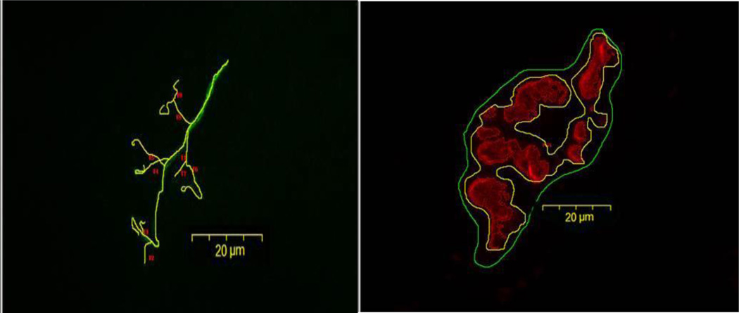
Figure 2.
Representative image of tracings used to quantify morphological aspects of neuromuscular junction from aged, control plantaris muscle. The left panel depicts tracings used to quantify presynaptic nerve terminal branches. The right panel illustrates tracings (both manually drawn and generated by software) used to quantify postsynaptic ACh receptors. Original magnification of × 1,000 (scale bar = 20 µm).

