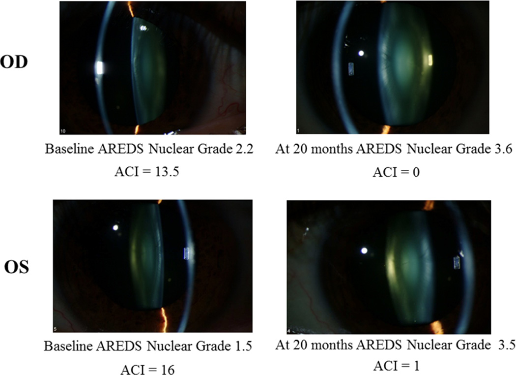
Figure 4.
Nuclear cataract progression in a patient with corresponding decrease in ACI. The upper two figures are for the right eye of a 43 year old patient, at baseline and 20 months later, and the lower two figures show the left eye of the same patient, at baseline and 20 months later. As the nuclear cataract progressed, the ACI decreased.

