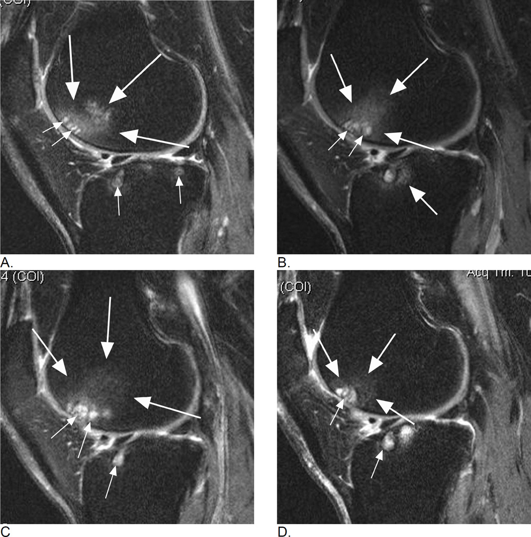
Figure 1.
Longitudinal assessment of bone marrow lesions (BML) in the lateral tibio-femoral and patello-femoral compartments. A. Baseline sagittal intermediate-weighted fat-suppressed MRI shows a grade 2 MOAKS / grade 3 WORMS BML in the anterior lateral femur displaying high-signal intensity, comprised of an ill-defined (edema-like) component (large arrows) and a well-defined cystic component (small arrows). In addition, there are small (MOAKS/WORMS grade 1) cystic BMLs in the subchondral anterior and posterior lateral tibia (small arrows). B. Follow-up MRI 1 year later shows slight decrease of overall lesion size (within-grade change for MOAKS, and change from grade 3 to grade 2 for WORMS) in the femur (large arrows) but increase of size of femoral cystic component (small arrows). While WORMS differentiates diffuse or ill-defined component from cystic component of BML as two distinct lesions, MOAKS takes both into account as one lesion and thus, would regard this as an overall decrease in lesion size while WORMS would score the increase in size of cystic portion as within-grade change. Note regression of cystic lesion in the posterior lateral tibia and increase of ill-defined (edema-like) portion of BML in the anterior lateral tibia now to a grade 2 lesion in MOAKS and WORMS (arrow). C. Further follow-up 1 year later, shows increase in overall lesion size in the anterior lateral femur (large arrows - now defined as grade 3 by both, MOAKS and WORMS). No relevant change in cystic portion of femoral and tibial BMLs in comparison to the previous visit is seen (small arrows). However, the ill-defined (edema-like) portion of the lesion in the anterior lateral tibia disappeared. D. 3 years after baseline, decrease in overall femoral lesion size (large arrows) is observed (now grade 2 by MOAKS and WORMS) while size of cystic portions of femoral and tibial BMLs remains stable (small arrows). However, there is progression of ill-defined portion of tibial BML (from grade 1 to grade 2 in the central lateral tibia for overall lesions size by MOAKS, and grade 0 to 1 for ill-defined BML only in WORMS).

