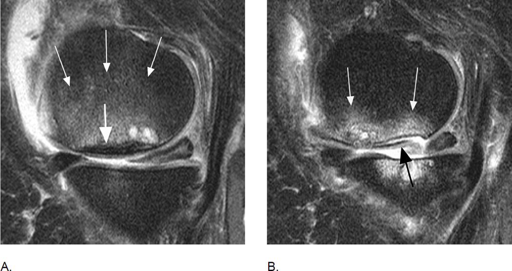
Figure 10.
Differential diagnosis of BMLs. A. Sagittal intermediate-weighted fat suppressed image shows a large grade 3 (MOAKS and WORMS) edema-like BML in the central medial femur (small arrows). Note diffuse subchondral hypointensity of 4 mm thickness representing a subchondral fracture in addition to the BML (large arrow). Further, there is a cystic portion adjacent to posterior aspect of the fracture line. B. 12 months later there is evidence of an osteochondral fracture with partial delamination of an osteochondral fragment (black arrow) and beginning deformity and collapse of the articular surface. Accompanying BML has decreased in size to a grade 2 (MOAKS) / grade 3 (WORMS) lesion (white arrows). In addition, at baseline there is a small central medial tibial BML (WORMS and MOAKS 1) with preserved cartilage. At follow-up, the BML increased in size (WORMS and MOAKS 3) with extensive loss of central and posterior tibial cartilage. The posterior horn of the medial meniscus showed a normal shape at baseline with linear intrameniscal signal reaching the undersurface representing a horizontal-oblique tear. At follow-up the posterior horn of the medial meniscus exhibits partial maceration. Note that effusion-synovitis decreased in size from baseline to follow-up.
Thick areas of hypointensity representing subchondral fracture (also known as spontaneous osteonecrosis of the knee or SONK) have a negative prognosis in regard to structural joint preservation31. SONK is not scored specifically in any of the scoring systems.

