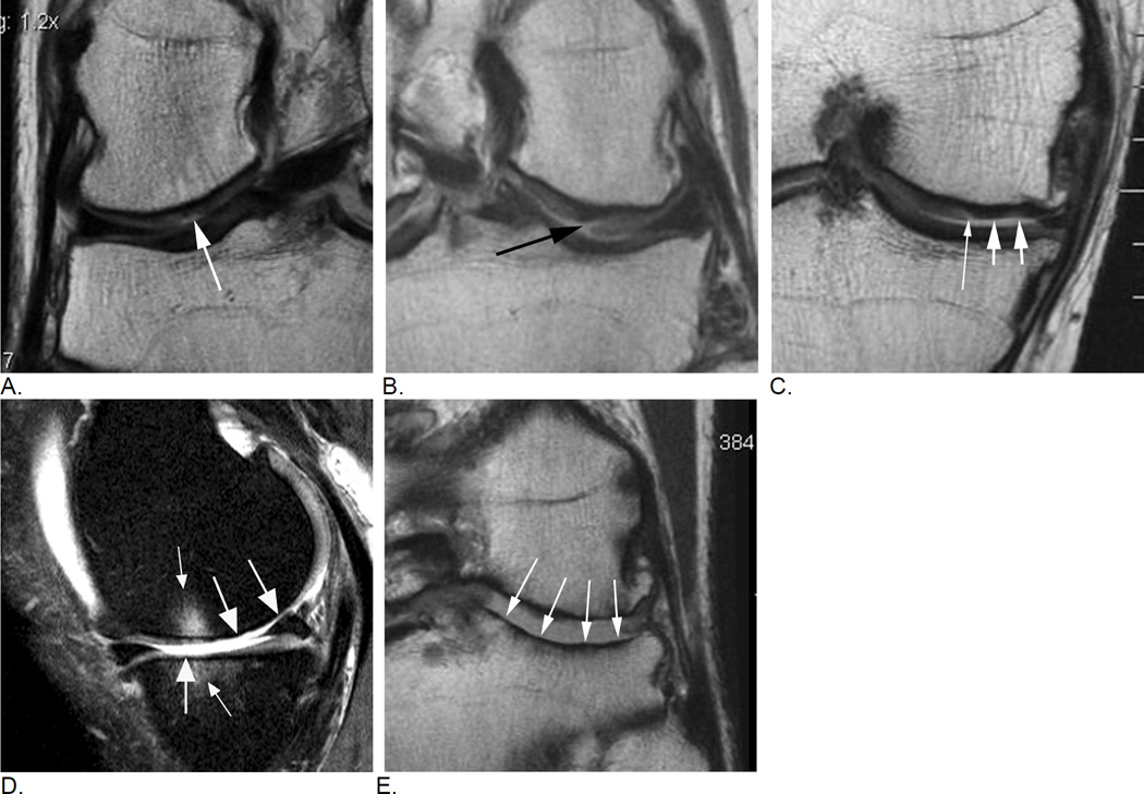
Figure 11.
Typical image examples for different types of cartilage damage. A. A focal superficial defect not reaching the subchondral plate is shown in this coronal intermediate-weighted MRI (arrow). Lesion will be coded as a grade 1.0 lesion in MOAKS (i.e. grade 1 - less than 10% in regard to area involvement, and grade 0 in regard to % of lesion that is full thickness damage) or as grade 2 in WORMS (defined as focal superficial defect less than 1 cm in maximum diameter). B. Coronal intermediate-weighted MRI shows a focal defect that reaches the subchondral plate and is consequently defined as a grade 1.1 lesion using MOAKS (less than 10% in area involvement, less than 10% of subregion that is full thickness damage). In WORMS this lesion would be scored as a 2.5 lesion. A 2.5 lesion is not a reflection of a within-grade coding but a distinct grade by itself. C. Another coronal intermediate-weighted MRI shows an example of a MOAKS 2.1 lesion, which is defined as superficial damage involving between 10% and 75% of subregion (short arrows) plus a full thickness component involving less than 10% of subregion (thin arrow). WORMS does not allow scoring of this lesion as it does not fulfill criteria of a focal defect only (i.e. a grade 2.5 lesion in WORMS), not of superficial damage only (grade 3 in WORMS) of diffuse full thickness damage (grade 5). Readers would have to decide which one of these grades applies best, which would be grade 3 lesion while ignoring the full thickness component. Whether a small full thickness component is of relevance in regard to clinical or structural outcome needs to be shown. D. Sagittal intermediate-weighted fat-suppressed MRI depicts diffuse full thickness cartilage damage in the central subregion of the medial femur and the central medial tibia representing grade 2.2. lesion in MOAKS, and grade 5 lesions in WORMS (large arrows). There are associated subchondral bone marrow lesions visualized as ill-defined hyperintense areas (small arrows). E. Another example shows extensive full thickness cartilage damage of the central lateral tibia (arrows) qualifying as a grade 3.3. lesion in MOAKS and a grade 6 lesion in WORMS. In addition, there are marked articular surface contour alterations of the tibia termed bone attrition, a feature only assessed in WORMS but not MOAKS.

