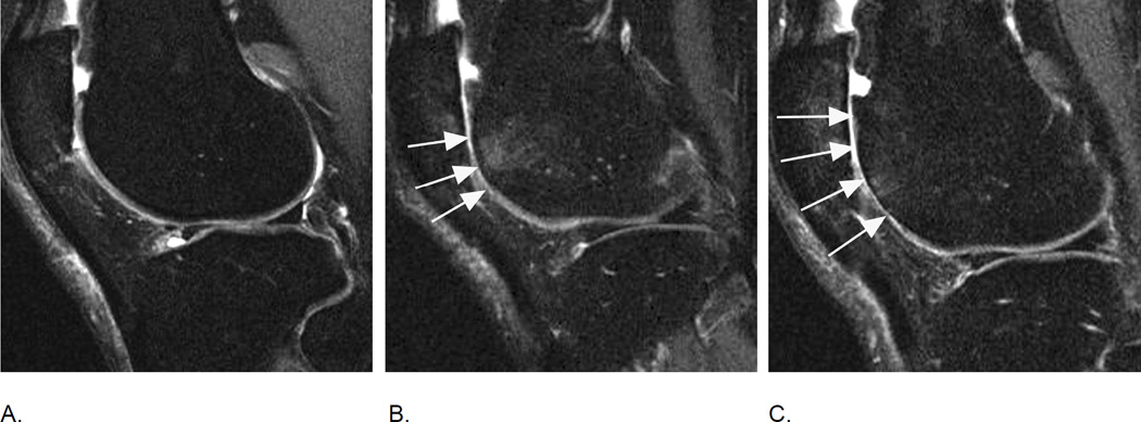
Figure 12.
Evolution of cartilage damage over time. A. Baseline fat-suppressed intermediate-weighted MRI shows an intact articular cartilage surface in the anterior lateral femur. There is diffuse full thickness cartilage damage at the patella (WORMS 6, MOAKS 3.3). B. 12 months later areas of partial and full thickness cartilage damage in the anterior lateral femur are observed that will be graded as a MOAKS 2.2 lesion (10–75% of subregion with any cartilage loss, 10–75% of subregion with full thickness cartilage loss) and a grade 5 lesion using WORMS (arrows). C. Another 12 months later there is definite increase in area extent of lesion (arrows). Using MOAKS this lesion would now represent a grade 3.2 lesion (> 75% of subregion with any cartilage loss, 10–75% of subregions showing full thickness loss), in WORMS it would qualify as a grade 6 lesion (more than 75% of subregion affected by full thickness cartilage loss), although there is still some cartilage preserved especially towards the more central part of the subregion. Also there is an anterior lateral femoral BML at the first follow-up visit (WORMS grade 2, MOAKS grade 1) which shows decrease in size 12 month later (WORMS and MOAKS grade 1).

