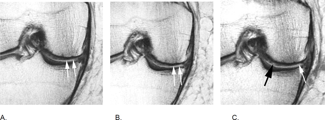
Figure 13.
Within-grade cartilage assessment. A. Baseline coronal proton density-weighted MRI shows superficial cartilage damage (arrows) qualifying as a MOAKS grade 2.0 lesion (10–75% of subregion affected by any cartilage loss with 0% of subregion being affected by full thickness damage), representing a WORMS grade 3 lesion (multiple areas of partial-thickness defects intermixed with areas of normal thickness, or a defect wider than 1 cm but <75% of the region). B. 12 months later, there is subtle but definite increase in cartilage loss (arrows) defined as within-grade change in both WORMS and MOAKS. C. At 24 months follow up there is further discrete increase in superficial cartilage damage (white arrow) defined as within-grade change. In addition there is an incident small full thickness defect more centrally (black arrow), which defines the lesion as a grade 2.1 using MOAKS. In WORMS lesion cannot be specifically graded and will likely be coded as a grade 3 lesion not taking into account the full thickness component.

