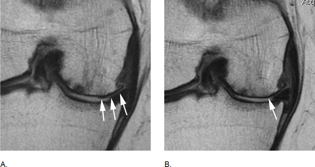
Figure 14.
Progression of cartilage damage in advanced disease. A. Baseline coronal proton density-weighted MRI shows diffuse full thickness cartilage loss (grade 3.3 MOAKS, grade 6 WORMS) in the central subregion of the medial femur. There is still cartilage remaining at the periphery of the joint (arrows). B. 12 months later there is definite progression of cartilage loss qualifying as within grade change (arrow). Although counter-intuitive from a clinical perspective, knees with advanced disease including radiographic stages grade 4 according to the Kellgren-Lawrence classification show the most rapid progression of cartilage loss32. Note that by the introduction of within-grade change the limitation of ceiling effects in ordinal scoring was overcome as even a grade 6 WORMS/ 3.3 MOAKS lesion may still progress and such progress may be coded as within-grade change.

