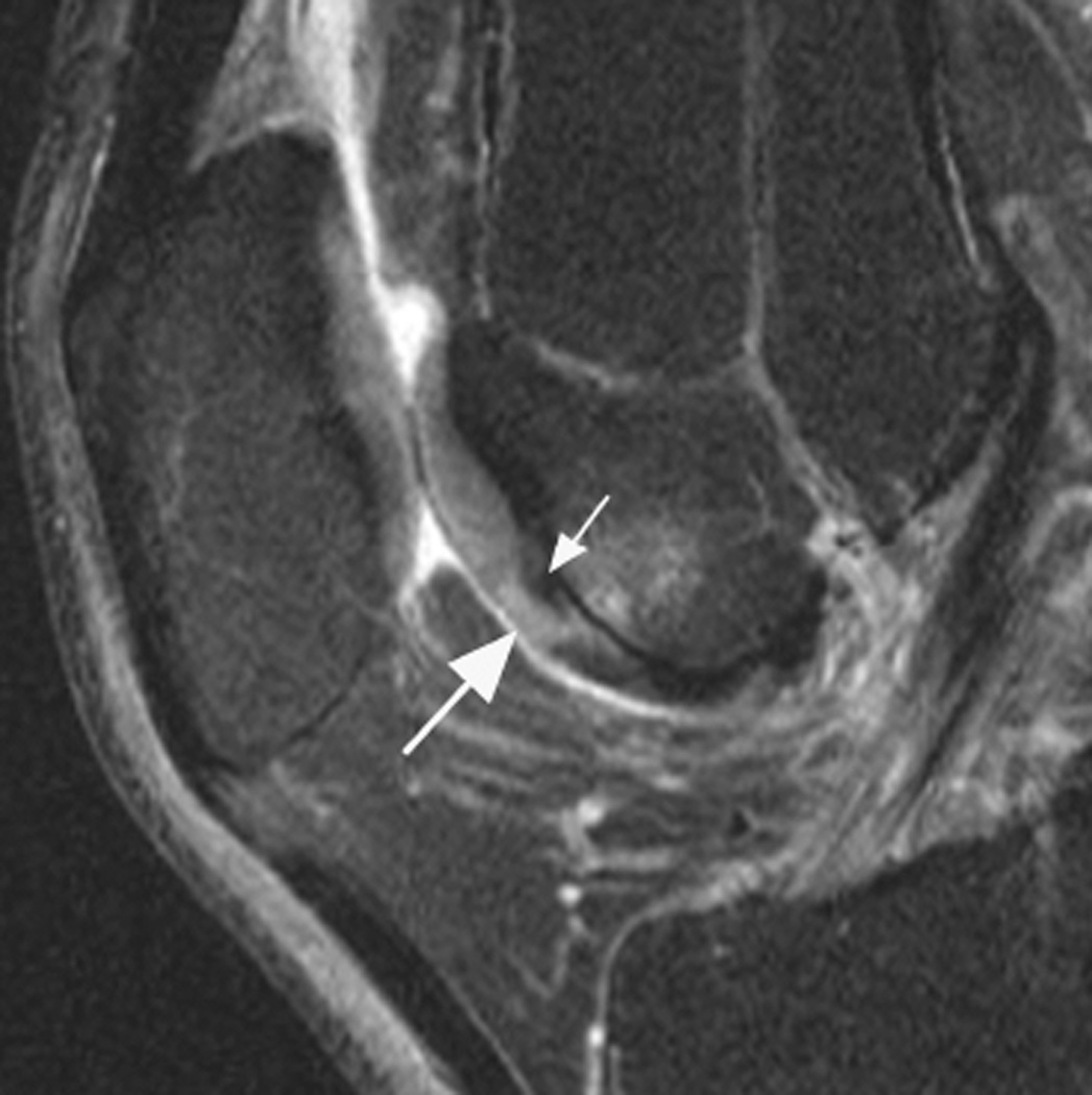
Figure 15.
Intrachondral signal change. A. Sagittal intermediate-weighted fat-suppressed MRI shows an intact cartilage surface without morphologic defects. There is an intrachondral hyperintensity signal change that is scored in WORMS as a grade 1.0 lesion but not coded in MOAKS as the clinical relevance of intrasubstance signal changes is unclear and dependent on MRI sequence applied. In addition, and particularly in the area of the femoral trochlea, so-called magic angle artifacts that occur at 55° to the main magnetic field (B0) may result in intracartilaginous signal alterations. In addition, image shows a discrete hypointense signal change that likely represents intrachondral calcification, which is not coded in either MOAKS or WORMS (small arrow). Note subchondral BML subjacent to the cartilage signal abnormality grade 1 at both WORMS and MOAKS

