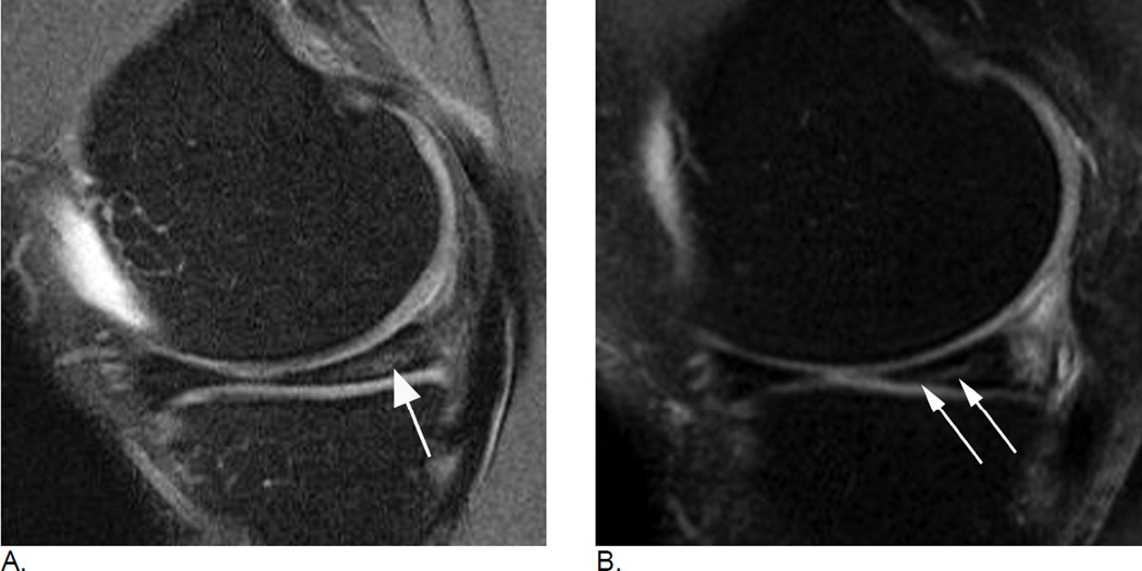
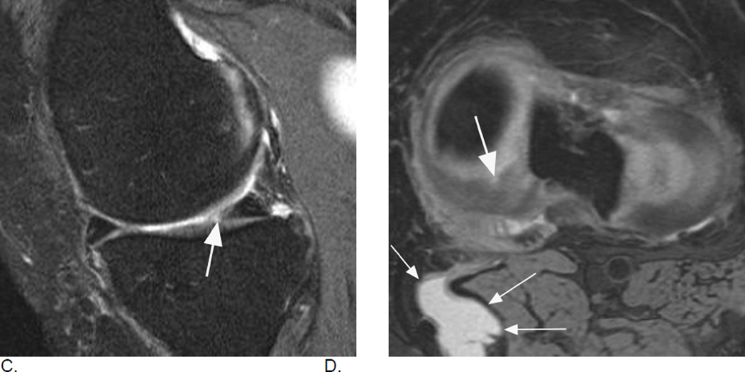
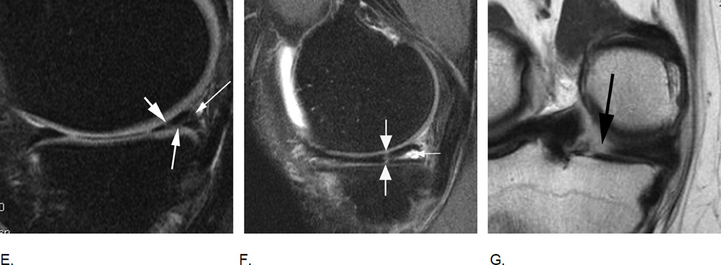
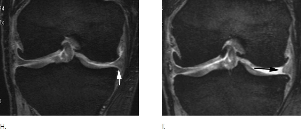
Figure 16.
Different types of meniscal damage. While WORMS uses a 0 to 4 scale to grade meniscal damage, the MOAKS systems applies a more complex and more comprehensive assessment of meniscal pathology including meniscal signal, different tear types, and types of meniscal substance loss / maceration. A. Sagittal intermediate-weighted fat-suppressed MRI depicts intramensical high signal in the posterior horn of the medial meniscus that does not reach the meniscal surface (arrow). Intrameniscal signal is only scored in the MOAKS system and is considered to represent mucoid degeneration of the meniscal fibrocartilage. Clinical relevance of meniscal signal is being discussed controversially. B. Example of a horizontal-oblique tear reaching the meniscal undersurface (arrows). This tear type is part of the spectrum of structural MRI manifestations of OA. A tear is defined on MRI as linear meniscal signal that reaches the meniscal surface on at least two consecutive slices33. The present example would be scored as a grade 1 lesion (parrot-beak) in the WORMS scale, while MOAKS uses a descriptive coding of meniscal pathology.
C. and D. Example of a radial tear. A radial tear involves the so-called “white”, non-vascularized inner zone of the meniscus. A small substance defect is seen on the sagittal intermediate-weighted fat suppressed image (arrow in C) that is confirmed and well-visualized on the corresponding high resolution axial dual-echo at steady-state (DESS) image in D (large arrow). In addition, there is a popliteal (Baker’s) cyst in the typical medial location between the tendons of the medial gastrocnemius and semimembranosus (small arrows).
E. Sagittal intermediate-weighted fat-suppressed MRI shows a complex meniscal tear of the posterior horn of the medial meniscus that reaches the posterior margin (thin arrow), the femoral (short arrow) and the tibial surfaces (intermediate arrow). Such a tear would be scored as a grade 2 lesion using the WORMS system. F. Another example of a complex tear shows a vertical tear of the medial posterior horn (large arrows) and in addition a horizontal tear communicating with an intrameniscal cyst (small arrow). Meniscal cysts are scored in MOAKS but not WORMS. G. Radial meniscal root tears are rare but clinically very relevant as these are associated with fast progression of cartilage loss. They are often associated with significant meniscal extrusion and hypertrophy. Coronal intermediate-weighted MRI shows a radial meniscal root tear of the medial posterior horn, which is visualized as a gap in the posterior central part of the meniscus (arrow). Root tears are coded in MOAKS, in WORMS these would be recorded as a grades 2 or 3 although not specifically mentioned in the scoring system.
H. Meniscal maceration is commonly observed in OA knees. Baseline coronal dual echo at stead state (DESS) image shows a normal body of the medial meniscus without evidence of a tear of substance loss but little extrusion (grade 1 MOAKS) I. Two years later, there is evidence of substance loss in concerning the central part of the body region (also referred to as the “white zone”). This is finding is also termed partial meniscal maceration.
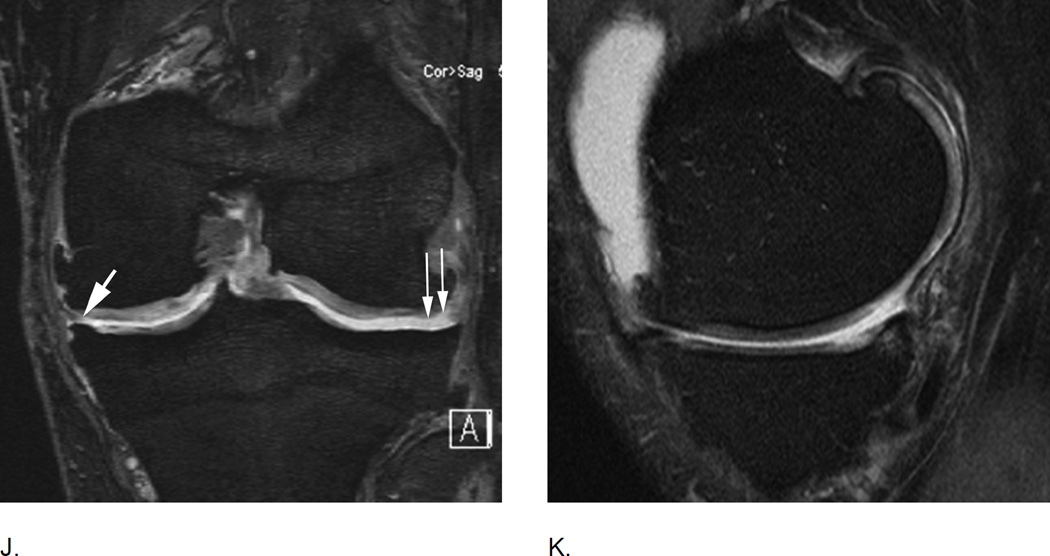
J. Coronal dual echo at steady state (DESS) image shows complete missing body of the medial meniscus (small thin arrows) consistent with complete maceration. There is advanced partial maceration laterally (short thick arrow). K. Corresponding sagittal intermediate-weighted fat suppressed image confirms complete substance loss of the medial meniscus likely due to meniscectomy. Non surgically induced maceration and surgical removal of the meniscus can not be differentiated unless there are other imaging signs that give evidence of prior surgery.

