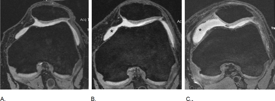
Figure 18.
MRI of markers of inflammation in OA. Fluid sensitive sequences are capable of delineating intraarticular joint fluid. However, a distinction between true joint effusion and synovial thickening is not possible as both are visualized as hyperintense signal within the joint cavity. For this reason the term effusion-synovitis has been introduced34, which is scored based on the distension of the joint capsule for both systems, WORMS and MOAKS, and is graded collectively from 0 to 3 in terms of the estimated maximal distention of the synovial cavity with 0=normal, grade 1=<33% of maximum potential distention grade 2=33%–66% of maximum potential distention and grade 3=>66% of maximum potential distention. Axial dual-echo at steady-state (DESS) MR images show A. grade 1 effusion-synovitis, B. grade 2 effusion-synovitis (asterisk) with medial patellar cartilage damage and C. grade 3 effusion-synovitis (asterisk).

