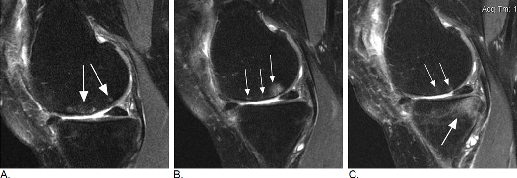
Figure 2.
Longitudinal BML assessment. Relevance of lesional vs. subregional scoring. A. Baseline sagittal intermediate-weighted fat-suppressed MRI shows two distinct ill-defined edema-like lesions in the central subregion of the medial femur (arrows). Overall lesion size in subregion qualifies as a grade 1 MOAKS / grade 2 WORMS lesion. B. Follow-up MRI one year later shows within-grade increase in overall subregional lesion size in the same subregion. In contrast to image A., now there are three distinct lesions (arrows). The single anterior lesion has split into two lesions with a decrease in lesion size, while the previous posterior lesion shows now an increase in lesion size. Note diffuse femoral cartilage loss in addition. C. Two-year follow-up MRI shows a decrease in overall femoral BML size with now a total subregional score of 1 using WORMS and MOAKS. There are two distinct lesions now with the most anterior lesion from image B. showing complete regression (small arrows). No cystic portion of lesions is observed. There is a large (grade 3 WORMS and MOAKS) incident lesion in the posterior medial tibia (large arrow). Without the clinical context posterior lesion can not be further characterized as traumatic bone contusions e.g. due to an anterior cruciate ligament tear may exhibit identical image morphology (reticular pattern with very diffuse borders atypical for OA-associated BMLs). Note further that articular cartilage in the posterior tibia is intact, which may further suggest a possible traumatic origin as OA-related BMLs are commonly associated with cartilage damage.

