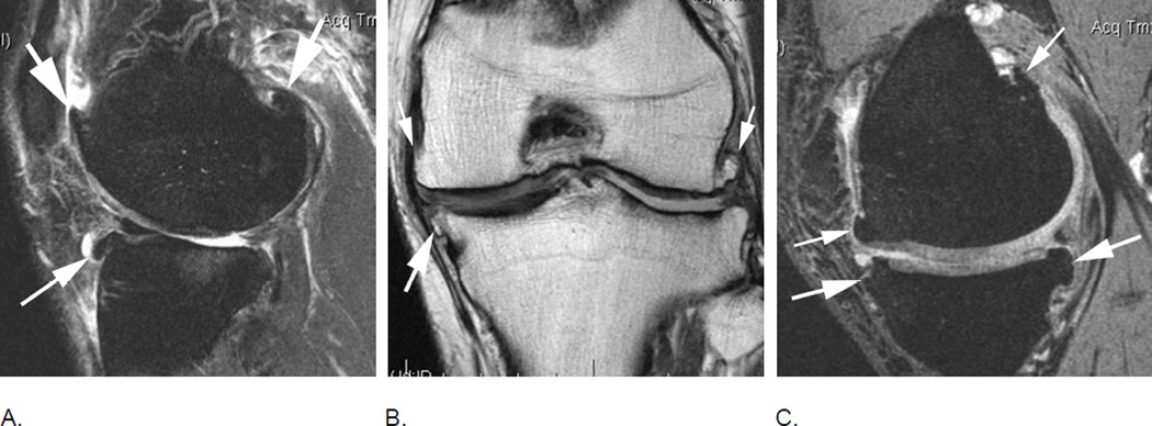
Figure 21.
Osteophytes are one of the hallmark features of OA on imaging and part of the disease definition on X-rays. While WORMS uses a complex approach of osteophytes scoring on a 0–7 scale at 16 articular anatomical locations, MOAKS applies a somewhat simplified scheme on a 0–3 scale at only 12 different locations omitting the scores of the anterior and posterior medial and lateral tibia. A. Sagittal fat-suppressed intermediate-weighted image of the lateral tibio-femoral compartment shows a moderate sized MOAKS grade 2 / WORMS grade 4 osteophyte at the anterior femur, a MOAKS grade 3 / WORMS grade 5 osteophyte at the posterior femur (short arrows) and a WORMS grade 5 osteophyte (long arrow) at the anterior lateral tibia (location not considered in MOAKS). Note diffuse cartilage loss at the central and posterior lateral tibia and femur and subchondral BML of the lateral tibial plateau, with moderate bone remodeling (attrition grade 2 in WORMS). B. Marginal osteophytes in the coronal plane are similarly considered in MOAKS and WORMS. Example shows femoral osteophytes (small arrows – MOAKS grade 2 / WORMS grade 4 medial; MOAKS grade 3 / WORMS grade 6 lateral) and a moderate osteophyte at the medial tibia (large arrow – MOAKS grade 2 / WORMS grade 4). There is diffuse cartilage loss at the central lateral tibial and femur with moderate lateral tibial plateau remodeling (attrition). C. Sagittal dual-echo at steady-state (DESS) MRI of the medial tibio-femoral compartment shows moderate-sized (MOAKS grade 2 / WORMS grade 3) osteophytes at the anterior and posterior medial femur (small arrows). At the tibia there is a tiny anterior osteophyte (WORMS grade 1) and a moderate-to-large sized posterior osteophyte (WORMS grade 5). Tibial locations are not scored in the sagittal plane using MOAKS.

