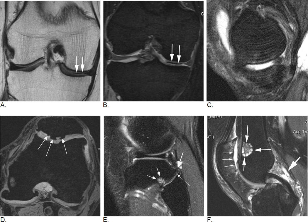
Figure 24.
Readers will encounter incidental findings and artifacts that either are not covered by the scoring systems or may impair image assessment. Any reader should have profound knowledge of MRI artifacts in order to distinguish these artifacts from potentially clinically relevant findings. A. Coronal intermediate-weighted (spin echo) MRI shows a hypointense line within the medial tibio-femoral joint space extending to and not to be differentiated from the body of the medial meniscus (arrows). B. Coronal DESS (gradient echo) MRI shows that hypointensity is a distinct structure not in relation to the meniscus (arrows). Finding represents a susceptibility artifact due to vacuum phenomenon, a common finding in knee OA that potentially may impair especially quantitative image assessment38. C. Motion. Sagittal intermediate-weighted fat-suppressed image shows repetitive hyperintense lines through the femoral and tibial subchondral bone mirroring the articular surface contour (arrows). Finding represents a motion artifact (also called zebra artifact) due to knee movement during the MRI acquisition. This artifact primarily impairs assessment of the cartilage and subchondral bone but may also mimic a meniscal tear. D. Axial DESS MRI depicts hypointense changes within the patellar cartilage that represent intrachondral osteophytes of unknown clinical relevance (arrows). These osteophytes commonly do not alter the cartilage surface and may only be detected on imaging but not arthroscopy. Neither scoring system assesses intrachondral osteophytes. E. Tibio-fibular OA is not scored in MOAKS or WORMS. Sagittal intermediate-weighted fat suppressed image shows characteristic signs of OA in this example including a tibial osteophyte (large arrow), joint space narrowing (long thin arrow) and a tibial subchondral bone marrow lesion (short small arrows). Especially in the absence of features of tibio-femoral OA, tibio-fibular OA may explain clinical symptoms such as lateral knee pain. F. An ovoid, lobulated hyperintense lesion is seen in the anterior aspect of the metaphyseal femur (intermediate-sized arrows). The chondroid matrix (salt and pepper-appearance on MRI) of the lesion is characteristic for a typical enchondroma, a finding of no or only little clinical relevance. Note additional features of OA such as diffuse full thickness cartilage loss of the lateral patella (small arrows), and marked effusion-synovitis posterior to the posterior cruciate ligament (large arrow), the commonest location of synovitis in the OA joint39.

