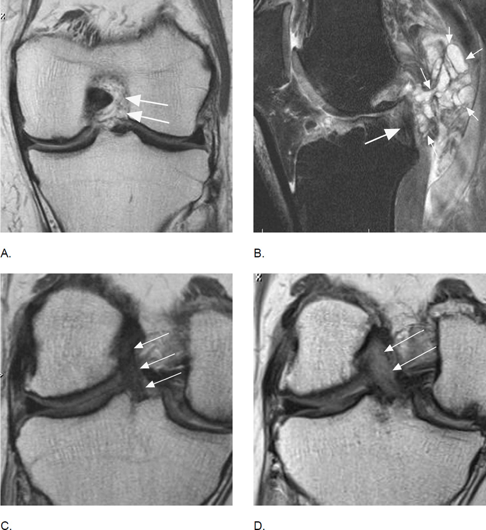
Figure 25.
Anterior cruciate ligament tears are associated with osteoarthritis longitudinally but may also be part of the disease process without a history of trauma. A. Coronal intermediate-weighted MRI shows absence of the ACL. Only intraarticular, extrasynovial fat and no ligamentous fibers are seen at the typical location of the ACL (arrows). B. Another example shows a partial tear of the posterior cruciate ligament (PCL) with hyperintense signal changes and thinning of the ligament (large arrow). In addition there is a large multi-lobulated ganglion cyst adjacent to the PCL (small arrows) that may be a source of symptoms. Further, note diffuse fissuring of the cartilage of the anterior lateral femur. C. Baseline coronal image shows a normal course, signal intensity and thickness of the ACL (arrows). D. Corresponding follow-up image 24 months later shows marked thickening and diffuse hyperintensity of the ligament representing mucoid degeneration, a finding not covered by either scoring system that may potentially be of clinical relevance40. In addition, note diffuse cartilage loss at the medial central femur and tibia, which has progressed at follow-up with an incident subchondral femoral cyst.

