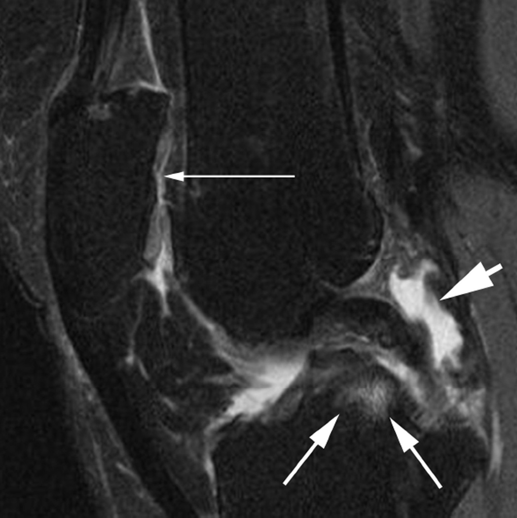
Figure 5.
BML in subspinous region. BMLs are scored in 15 articular subregions in both MOAKS and WORMS systems. While 14 of these subregions are in a subchondral location, one subregion is delineated by the tibial spines and is not associated with adjacent cartilage lesions but rather a result of traction to the cruciate ligaments that insert close to the tibial spines. Thus, when analyzing associations between subchondral BMLs and cartilage loss or clinical manifestations of disease, the subspinous BMLs are commonly excluded. Image example (sagittal fat-suppressed intermediate-weighted MRI) shows a grade 2 subspinous BML (intermediate sized arrows). Note additional partial thickness cartilage damage (grade 2.0 MOAKS, grade 3 WORMS) at the lateral patella (long arrow). In addition there is effusion-synovitis posterior to the posterior cruciate ligament, a common site of inflammatory disease manifestations in OA (short arrow).

