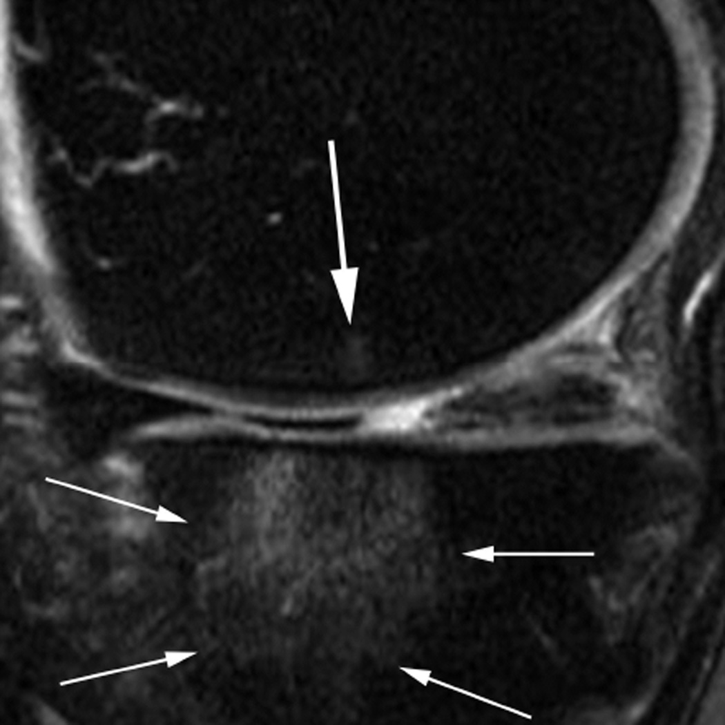
Figure 6.
Subregional BML assessment. Sagittal intermediate-weighted fat-suppressed image shows a large tibial edema-like BML that encompasses the anterior and central medial tibial subregions. Although radiologically only one lesion is observed (thin arrows), from a scoring perspective the lesion will be coded in two distinct subregions, a grade 3 (WORMS) / grade 2 (MOAKS) lesion the central tibia and a grade 2 (WORMS) / grade 1 (MOAKS and WORMS) lesion in the anterior tibia. In addition there is a small grade 1 (MOAKS and WORMS) edema-like lesion in the central medial femur (large arrow).

