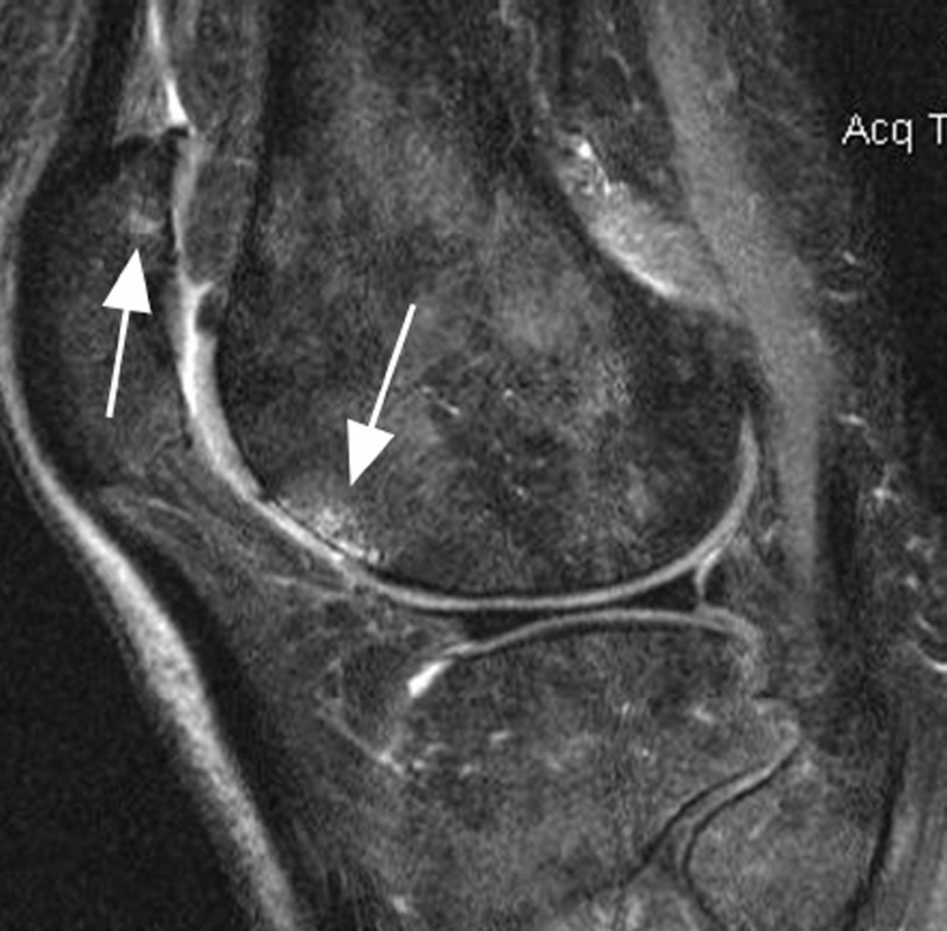
Figure 7.
Differential diagnosis of BMLs. In cases of marrow infiltration or reconversion BMLs may be difficult to distinguish as the bone marrow has a hyperintense appearance that may be identical to OA-related BMLs. Cystic portions of lesions can usually be detected with adequate sensitivity but ill-defined (edema-lile) portions of lesions may not be discernible from red marrow or marrow infiltration. This example (sagittal fat-suppressed intermediate-weighted MRI) shows diffuse hyperintense marrow reconversion (due to thalassemia) and grade 1 (in MOAKS and WORMS) BMLs at the anterior lateral femur and the lateral patella (arrows). Note that only the cyst-like portion of lesion can be detected in femoral BML due to marrow reconversion.

