Figure 8.
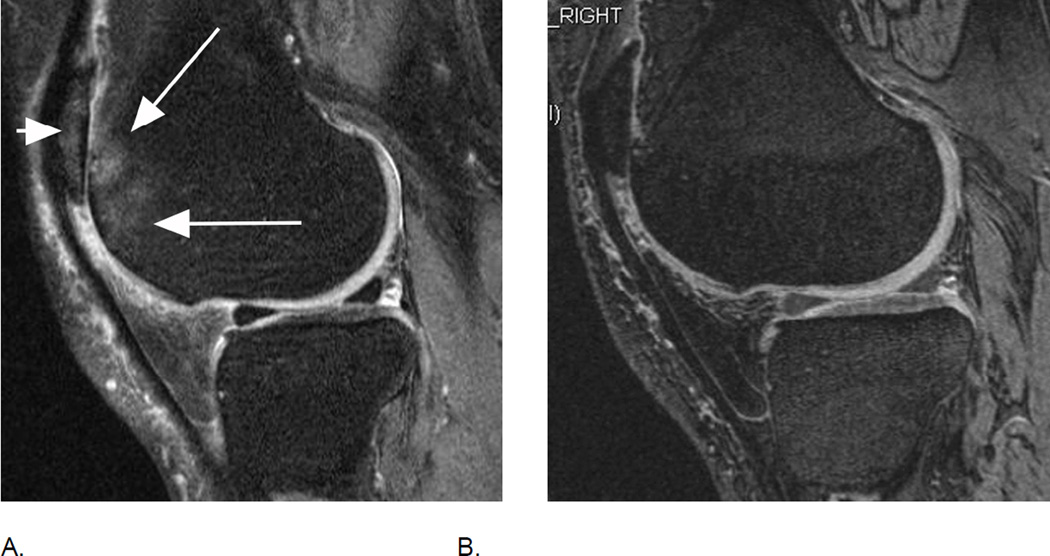
Relevance of sequence selection for BML assessment. A. Sagittal intermediate-weighted fat-suppressed MRI shows diffuse, ill-defined BML in the anterior lateral femur (long arrows), representing a grade 3 lesion in MOAKS and WORMS. There is also a BML at the lateral patella (short arrow), also coded grade 3 in MOAKS and WORMS. B. Corresponding dual-echo at steady-state (DESS) MRI does not allow differentiation of BMLs from normal marrow due to magnetic susceptibility, although DESS is a sequence with T2-weighted image characteristics. This low sensitivity for BML detection, especially for edema-like lesions, is common for all gradient echo sequences, including DESS.

