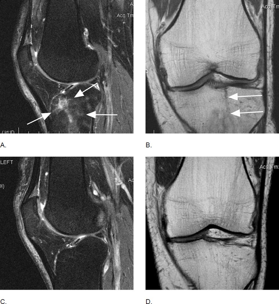
Figure 9.
Traumatic BMLs. Differentiation between traumatic and degenerative OA-related BMLs may be challenging22. This example allows a definite MRI-based diagnosis of a traumatic BML. A. Baseline sagittal fatsuppressed intermediate-weighted MRI shows a diffuse BML that is distant to the subchondral plate (arrows), which is a finding not seen in OA-related BMLs. B. Coronal proton density-weighted MRI shows a definite hypointense fracture line in the anterior tibia that confirms the traumatic origin of this finding (arrows). Follow- up images (C and D) 12 months later show complete resolution of lesion and fracture line, which is common for traumatic BMLs29,30.

