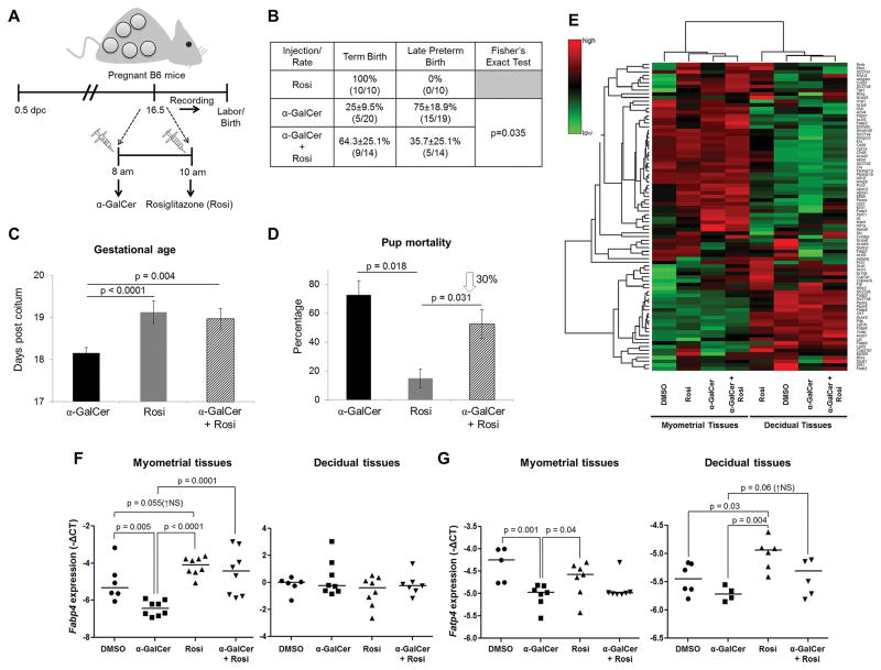
Figure 2. Rosiglitazone treatment reduces the rate of α-GalCer-induced late PTB by inducing PPARγ activation at the maternal-fetal interface.
(A) On 16.5 dpc, pregnant mice were i.v. injected with α-GalCer and treated shortly after with rosiglitazone (Rosi; s.c.) and video monitored (n=14). Control mice were s.c. injected with rosiglitazone alone (n=10). (B) The rate of term birth was defined as the percentage of dams delivering at 19.5±0.5 dpc among all births. The rate of late PTB was defined as the percentage of dams delivering between 18.0 and 18.5 dpc among all births. Data are represented as percentages ± 95% confidence interval. (C) Gestational age was calculated from the presence of the vaginal plug (0.5 dpc) until the observation of the first pup in the cage bedding. (D) The rate of pup mortality for each litter was defined as the proportion of born pups found dead out of the total litter size. (E) A heat map visualization of PPAR targets gene expression in myometrial and decidual tissues from dams i.v. injected with DMSO, α-GalCer, rosiglitazone, or α-GalCer + rosiglitazone. Data are from individual dams, n=4 each. (F) mRNA expression of Fabp4 in myometrial and decidual tissues. (G) mRNA expression of Fatp4 in myometrial and decidual tissues. Negative ΔCT values (F&G) were calculated using Actb as a reference gene. Data are from individual dams, n=6–8 each.

