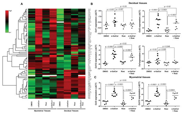
Figure 6. Administration of α-GalCer induces a pro-inflammatory microenvironment at the maternal-fetal interface that is partially attenuated by rosiglitazone.
(A) A heat map visualization of cytokine and chemokine gene expression in myometrial and decidual tissues from dams i.v. injected with DMSO, α-GalCer, rosiglitazone (Rosi) or α-GalCer + rosiglitazone. Data are from individual dams, n=4 each. (B) mRNA expression of Ccl1, Ccl2, Ccl12, and Tnf in decidual tissues. Negative ΔCt values were calculated using Actb as a reference gene. Data are from individual dams, n=6–8 each. (C) mRNA expression of Ccl2 and Ccl12 in myometrial tissues. Negative ΔCt values (B&C) were calculated using Actb as a reference gene. Data are from individual dams, n=6–8 each.

