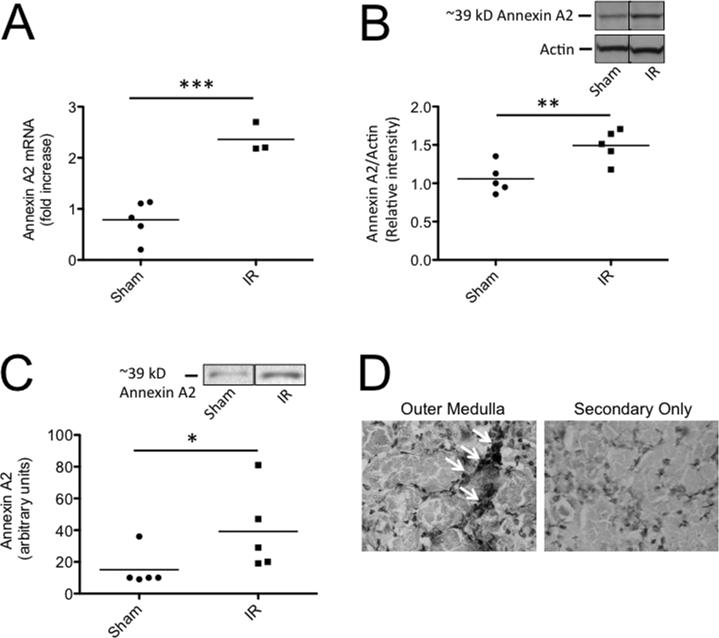
Figure 3. Annexin A2 is expressed in the kidney after ischemia/reperfusion.
C57BL/6 mice were subjected to renal ischemia and 24 hours of reperfusion. A) RNA was isolated from kidneys and the expression of annexin A2 was measured by RT-PCR. Annexin A2 expression was significantly increased after ischemia/reperfusion. B) Annexin A2 was detected in kidney lysates by Western blot analysis and was normalized to levels of actin. The relative intensity of annexin A2 protein in the two groups was compared by Student's T test, and annexin A2 protein levels were significantly increased after ischemia/reperfusion. Bands from two non-adjacent lanes are shown. C) Biotinylated mouse factor H was used to immunoprecipitate binding partners from kidney lysates, and the immunoprecipitated proteins were examined by Western blot analysis with an antibody for annexin A2. The abundance of annexin A2 immunoprecipitated from the kidneys was greater in post-ischemic kidneys than in sham treated kidneys. Bands from two non-adjacent lanes are shown. The groups were compared with Student's T tests. D) Immunohistochemistry of kidney sections for annexin A2 demonstrated peri-tubular localization of the protein in the outer medullae of post-ischemic mice. Original magnification 400X.

